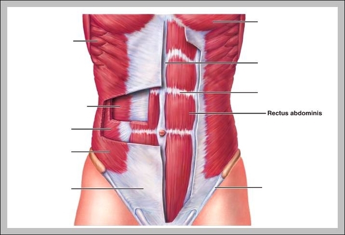
Rectus

This human anatomy diagram with labels depicts and explains the details and or parts of the Rectus. Human anatomy diagrams and charts show internal organs, body systems, cells, conditions, sickness and symptoms information and/or tips to ensure one lives in good health.

n. pl. rec·ti (-tī′) Any of various straight muscles, as of the abdomen, eye, neck, and thigh. Any of several straight muscles, such as the central vertical muscle on either side of the midline of the abdomen (the rectus abdominis), the rectus femoris on the front of the thigh or the four rectus muscles that move the eyeball.

“Recti” refers to your ab muscles called the “rectus abdominis.”. When the ab muscles move aside like this, the uterus, bowels, and other organs have only a thin band of connective tissue in front to hold them in place.

Rectus abdominis is formed by three or more transverse divisions above navel. This example is from Wikipedia and may be reused under a CC BY-SA license. This membrane is in relation in front with the rectus capitis anterior muscles, behind with the alar ligaments. This example is from Wikipedia and may be reused under a CC BY-SA license.

Rectus

rectus