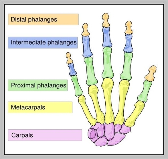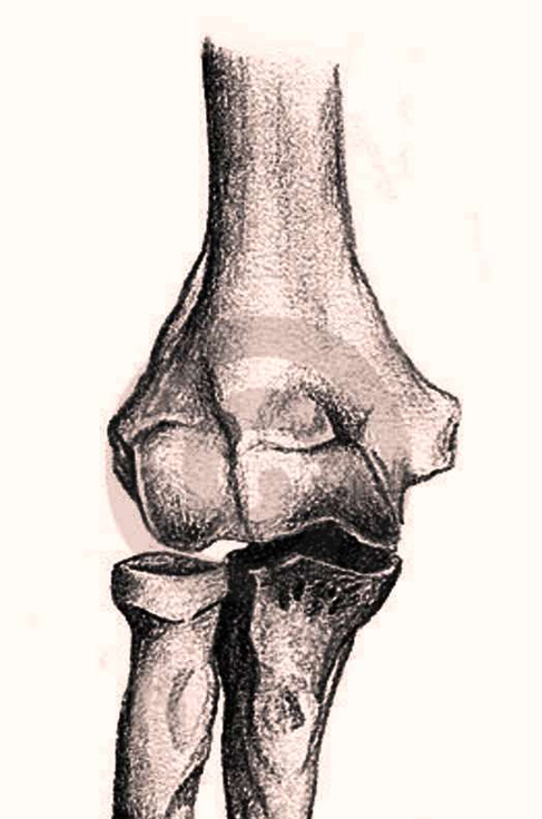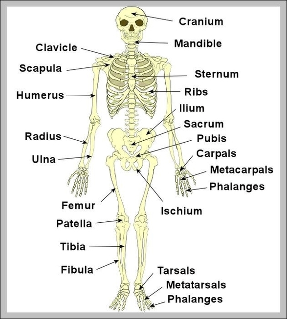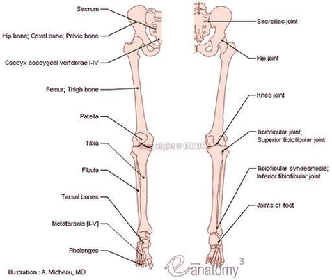
Arm Bones Diagram Image
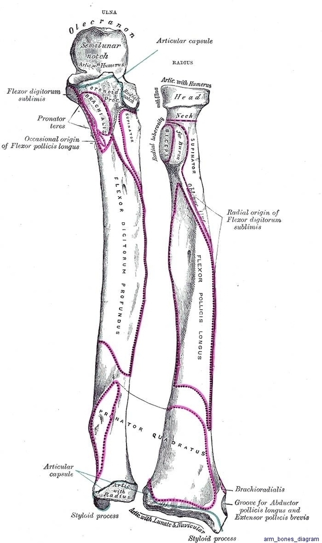
The human arm is divided into two main sections: the forearm and the arm itself. The forearm is the section located between the elbow and the wrist, while the arm represents the section from the shoulder to the elbow. The View Diagram Arm Bones Diagram Image