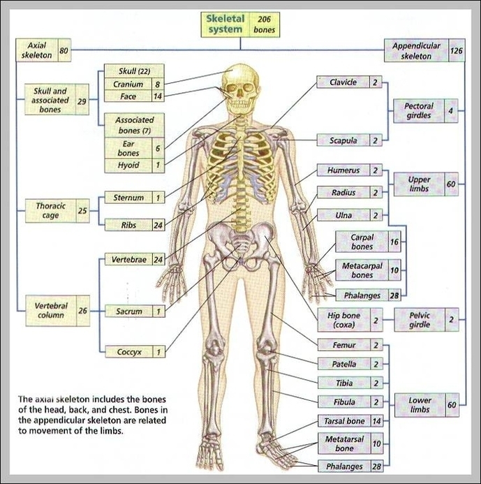
206 Bones Of The Body Diagram Image

This human anatomy diagram with labels depicts and explains the details and or parts of the 206 Bones Of The Body Diagram Image. Human anatomy diagrams and charts show internal organs, body systems, cells, conditions, sickness and symptoms information and/or tips to ensure one lives in good health.

Out of 206 bones, some bones are paired and each bone has different functionality. 206 Bones are classified into the following 4 types. Here is the complete list of all human bones.

SkullFig. 7.1(TE Art) Frontal bone Clavicle Maxilla Parietal bone Mandible Temporal bone Occipital bone Mandible Humerus Femur Tibia Calcaneus Fibula Ulna Radius Scapula Clavicle Scapula Vertebral column Pelvic girdle Patella Metacarpal bones Carpus Tarsus Thoracic cage Sternum Ribs Phalanges Metatarsal bones Phalanges Zygomatic bone

These bones are arranged into two major divisions: the axial skeleton and the appendicular skeleton. The axial skeleton runs along the body’s midline axis and is made up of 80 bones in the following regions:

206 Bones Of The Body Diagram Image

206 Bones Of The Body Diagram Image


Tags: , ,