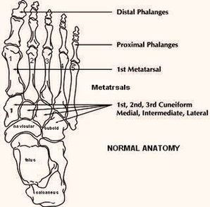
Foot Anatomy Image

This human anatomy diagram with labels depicts and explains the details and or parts of the Foot Anatomy Image. Human anatomy diagrams and charts show internal organs, body systems, cells, conditions, sickness and symptoms information and/or tips to ensure one lives in good health.

The anatomy of the foot. The foot contains a lot of moving parts – 26 bones, 33 joints and over 100 ligaments. The foot is divided into three sections – the forefoot, the midfoot and the hindfoot. The forefoot. This consists of five long metatarsal bones and five shorter bones that form the toes (phalanges).

2,416 human foot anatomy stock photos and images available, or start a new search to explore more stock photos and images.

Foot Definition The foot is a part of vertebrate anatomy which serves the purpose of supporting the animal’s weight and allowing for locomotion on land. In humans, the foot is one of the most complex structures in the body.

Foot Anatomy Image

Foot Anatomy Image


Tags: