Foot Anatomy Bones1 Image

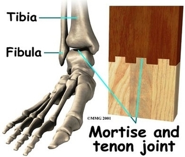
The anatomy of the foot. The foot contains a lot of moving parts – 26 bones, 33 joints and over 100 ligaments. The foot is divided into three sections – the forefoot, the midfoot and the hindfoot. The forefoot. This consists of five long metatarsal bones and five shorter bones that form the toes (phalanges).

The bones of the foot are organized into the tarsal bones, metatarsal bones, and phalanges. The foot begins at the lower end of the tibia and fibula, the two bones of the lower leg. At the base of those, a grouping of bones form the tarsals, which make up the ankle and upper portion of the foot.

Anatomy of the foot. Calcaneus (heel bone) Talus (ankle bone) Transverse tarsal joint. Navicular bone. Lateral cuneiform bone. Intermediate cuneiform bone. Medial cuneiform bone.

Foot Anatomy Bones1 Image Diagram - Chart - diagrams and charts with labels. This diagram depicts Foot Anatomy Bones1 Image

Foot Anatomy Bones1 Image