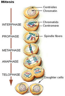
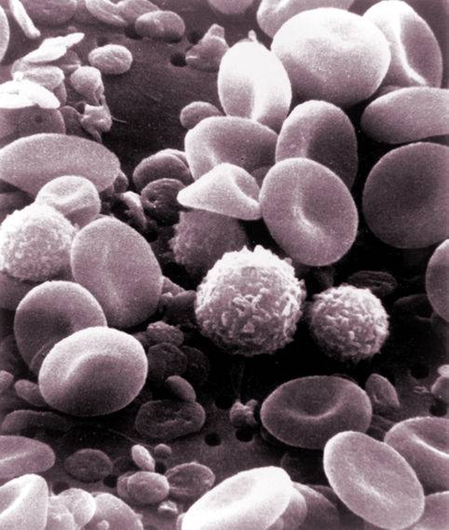
Hart Cell Cycle Diagram Image
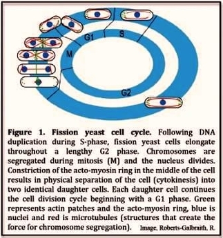
The phases are: 1. G1 (gap1) phase 2. S (synthesis) phase 3. G2 (gap 2) phase 4. M (mitosis) phase. Cell Cycle: Phase # 1. The Cardiac Cycle. The cardiac cycle includes all of the events that take place during View Diagram Hart Cell Cycle Diagram Image