Testis 2 Diagram Image

This human anatomy diagram with labels depicts and explains the details and or parts of the Testis 2 Diagram Image. Human anatomy diagrams and charts show internal organs, body systems, cells, conditions, sickness and symptoms information and/or tips to ensure one lives in good health.

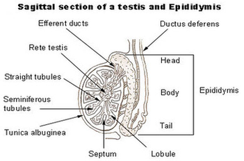
What are testes? The testes — also called testicles — are two oval-shaped organs in the male reproductive system. They’re contained in a sac of skin called the scrotum. The scrotum hangs outside the body in the front of the pelvic region near the upper thighs.

The testes (testicles) are male reproductive glands found in a saccular extension of the anterior abdominal wall called the scrotum. They are in ovoid shape, sized four to six centimeters in length. Testes develop retroperitoneally on the posterior abdominal wall and descend to scrotum before birth.

Each testis is divided by invaginations of the tunica albuginea that divide it into several hundred small segments called lobules. Each lobule contains several tightly coiled tubes called seminiferous tubules. The walls of the seminiferous tubules contain the germ cells, Sertoli cells, and Leydig cells that give the testes their function.

Testis 2 Diagram Image

Testis 2 Diagram Image


Tags: ,