Diagram Anatomy Of The Eye Image

This human anatomy diagram with labels depicts and explains the details and or parts of the Diagram Anatomy Of The Eye Image. Human anatomy diagrams and charts show internal organs, body systems, cells, conditions, sickness and symptoms information and/or tips to ensure one lives in good health.

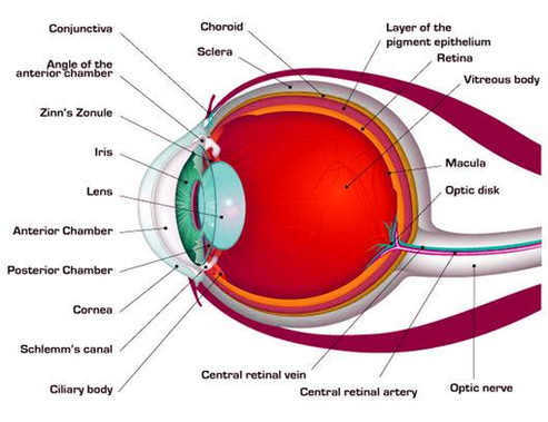
The human eye diagram is a visual depiction of the human eye. The following aspects are essential when constructing a human eye diagram . The conjunctiva is a thin, translucent layer of tissue that protects the front of the eyes, including the sclera and the eyelids inner surface.

The Anatomy of Human Eye The most complex sensory organs of the human body are the eyes. Every part of the human body is responsible for a specific action, from the muscles and tissues to the nerves and the blood vessels. The human eye consists of many muscles and tissues that join to form an approximately spherical structure.

In addition to the retina, other visual components such as the pupil, iris (colored part of the eye), and lens help focus light rays on the retina. The retina is the light-sensitive membrane covering the back of the eye. When light entering the eye hits the retina, special cells known as photoreceptors convert it into electrical signals. 1

Diagram Anatomy Of The Eye Image

Diagram Anatomy Of The Eye Image


Tags: