
Category Archives: Anatomy
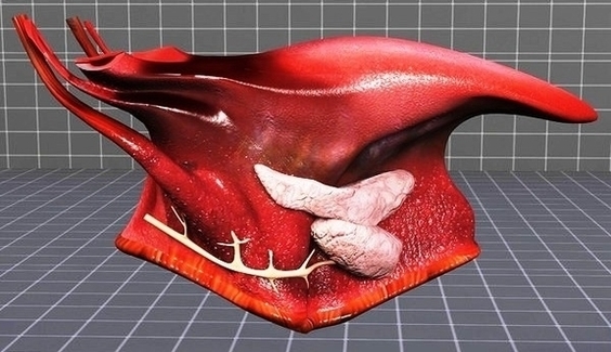
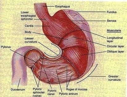
Stomach Anatomy Image
Cervical Disk Mri Image

Magnetic resonance imaging (MRI) is a safe, painless test that uses radio waves and energy from strong magnets to create detailed images of your body. A cervical MRI scans the soft tissues of your neck and cervical spine. The cervical View Diagram Cervical Disk Mri Image
Human Eye Diagram Image
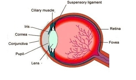
6,636 human eye diagram stock photos, vectors, and illustrations are available royalty-free. The Human Eye (Eyeball) Diagram, Parts and Pictures. The human eye consists of the eyeball, optic nerve, orbit and appendages (eyelids, extraocular muscles and lacrimal glands). While the View Diagram Human Eye Diagram Image
Anatomy Brain Nervous Fee Db Fa Ecffffclarge Image
Anatomy of the Brain The anatomy of the brain is complex due its intricate structure and function. This amazing organ acts as a control center by receiving, interpreting, and directing sensory information throughout the body. The brain and spinal cord View Diagram Anatomy Brain Nervous Fee Db Fa Ecffffclarge Image
Diabetes Diet Sheet Image
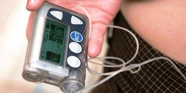
So an effort to use charts will be done. The way to get the optimal version of the diabetic diet chart is to consistently do every element in it. If you have done it regularly and don’t try to cheat View Diagram Diabetes Diet Sheet Image
Colorectal Cancer Diagram Image
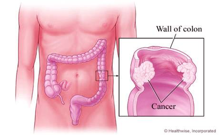
4,035 colorectal cancer stock photos and images available, or search for colon cancer screening or colonoscopy to find more great stock photos and pictures. Doctor goes over a patient”s x-ray, screening for colon cancer. There is no single cause of View Diagram Colorectal Cancer Diagram Image
Shoulder Anatomy Muscles Diagram Image
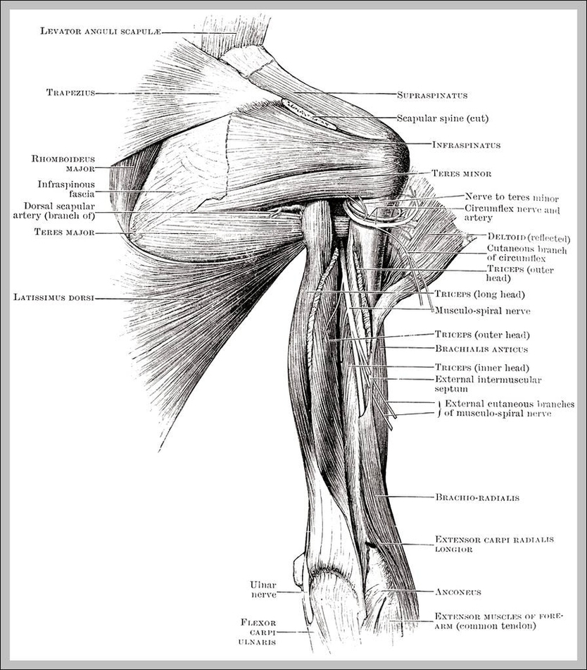
shoulder joint anatomy shoulder joint anatomy. Bones (Scapula, Humerus, Coracoid process, Acromion , Muscle (Biceps, Supraspinatus) and ligament (Coracohumeral) shoulder anatomy stock illustrations shoulder joint anatomy. 13,987 shoulder anatomy stock photos and images available, or search for shoulder anatomy muscles View Diagram Shoulder Anatomy Muscles Diagram Image
Ultrasoud Weeks Image

All about normal 10 week ultrasound. All about your normal 9 week ultrasound. Sharing is caring! Normal 13 week baby ultrasound. First let me tell you that by now you probably have all your first trimester regular ultrasounds done. The View Diagram Ultrasoud Weeks Image
Symptoms For Diabetes Image
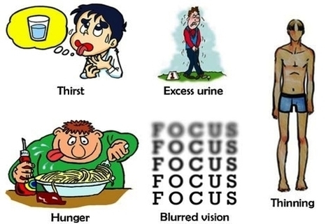
Diabetes Symptoms. 1 Urinate (pee) a lot, often at night. 2 Are very thirsty. 3 Lose weight without trying. 4 Are very hungry. 5 Have blurry vision. 6 Have numb or tingling hands or feet. 7 Feel very tired. 8 View Diagram Symptoms For Diabetes Image
Labeled Diagram Of Human Heart Image
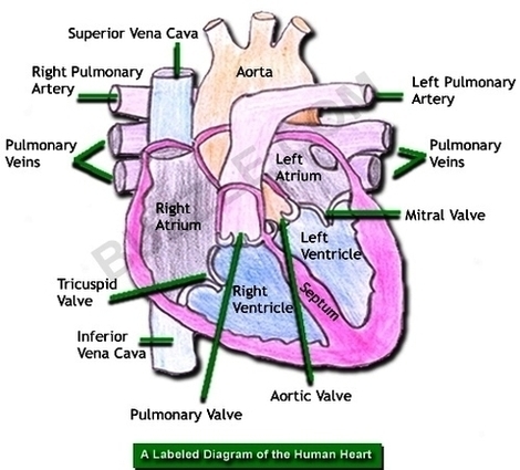
Human Heart Diagram Labeled. The human heart is an organ responsible for pumping blood through the body, moving the blood (which carries valuable oxygen) to all the tissues in the body. Without the heart, the tissues couldn’t get the oxygen View Diagram Labeled Diagram Of Human Heart Image
Dental Crown Tooth Before Image
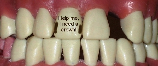
Dental crowns plays an important role in supporting and restoring the normal structure and appearance of damaged teeth. If any of your front teeth has suffered severe damage, broken or extensively decayed, a dental crown repair is usually recommended. However, View Diagram Dental Crown Tooth Before Image
Gestational Diabetes Symptoms Ukwhfqbi Image

Some pregnant women do notice subtle signs of gestational diabetes. The symptoms are similar to those of other forms of diabetes. But they’re also common symptoms in all pregnant women, so they’re easy to miss as the sign that something’s View Diagram Gestational Diabetes Symptoms Ukwhfqbi Image
Hiv Aids Chrt Photo Image

Adult Foot Fx Anatomya Image
Human Ear Diagram Image
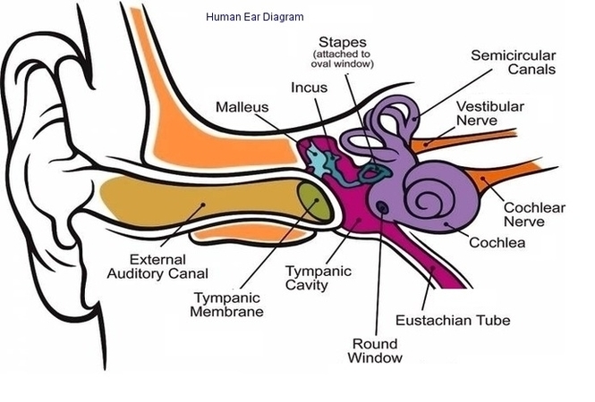
The Structure of Human Ear. Helix: It is the prominent outer rim of the external ear. Antihelix: It is the cartilage curve that is situated parallel to the helix. Crus of the Helix: It is the landmark of the outer View Diagram Human Ear Diagram Image
Motor Sensory Image
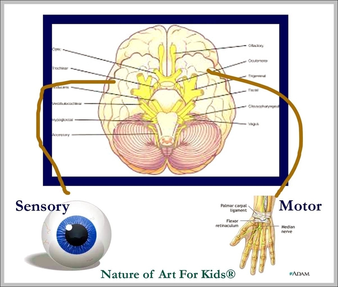
Motor imagery is the mental execution of a movement without any overt movement or without any peripheral (muscle) activation. It has been shown that motor imagery leads to the activation of the same brain areas as actual movement. Information from View Diagram Motor Sensory Image
Costovertebral Joints Anatomy Diagram Image

The costovertebral joints describe two groups of synovial plane joints which connect the proximal end of the ribs with their corresponding vertebrae, enclosing the thoracic cage from the posterior side. Precisely, these joints are described as; The costovertebral complex is View Diagram Costovertebral Joints Anatomy Diagram Image
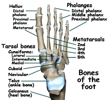
Footankle Bony Anat Image
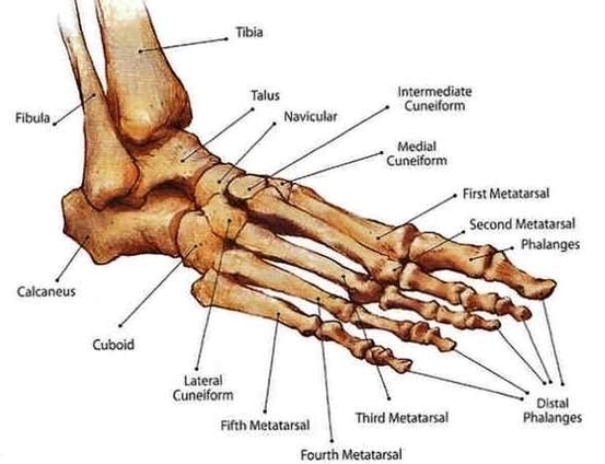
This article outlines the basic anatomy of the foot bones, along with some of the most common conditions affecting these bones. The human foot consists of 26 bones. These bones fall into three groups: the tarsal bones, metatarsal bones, and View Diagram Footankle Bony Anat Image
Aterial Pressure Fluctuations Image
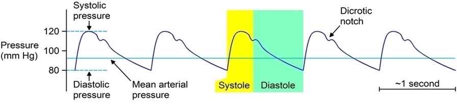
Arterial pressure results from the pressure exerted by the blood in the large arteries. Blood pressure depends on cardiac output and total peripheral resistance. Arterial pressure fluctuates with each heart beat, according to the pumping of the heart. Moreover, a View Diagram Aterial Pressure Fluctuations Image