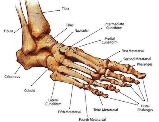
Footankle Bony Anat Image

This human anatomy diagram with labels depicts and explains the details and or parts of the Footankle Bony Anat Image. Human anatomy diagrams and charts show internal organs, body systems, cells, conditions, sickness and symptoms information and/or tips to ensure one lives in good health.

This article outlines the basic anatomy of the foot bones, along with some of the most common conditions affecting these bones. The human foot consists of 26 bones. These bones fall into three groups: the tarsal bones, metatarsal bones, and phalanges. The tarsal bones are a group of seven bones that make up the rear section of the foot.

Health Pages foot-ankle page provides pictures of the bones, ligaments, cartilages and tendons with explanation of common problems, treatment and function Skip to content Menu Menu Joints

For instance, while analogous bones do exist between the hand and the foot, such as the metatarsal bones and the phalanges (analogous to the metacarpal bones and phalanges of the hand) the bones of the feet are smaller, more sturdy in nature, and have limited motion when compared to the bones of the hand.

Footankle Bony Anat Image

Footankle Bony Anat Image


Tags: , ,