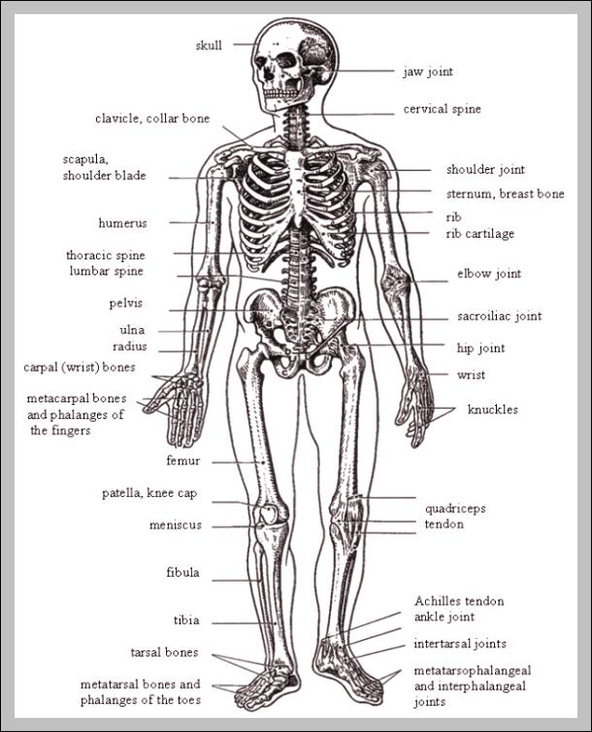
Pictures Of Bones In The Human Body

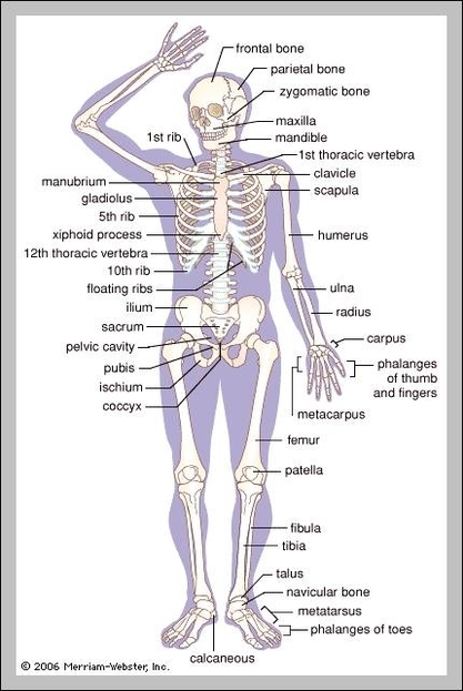
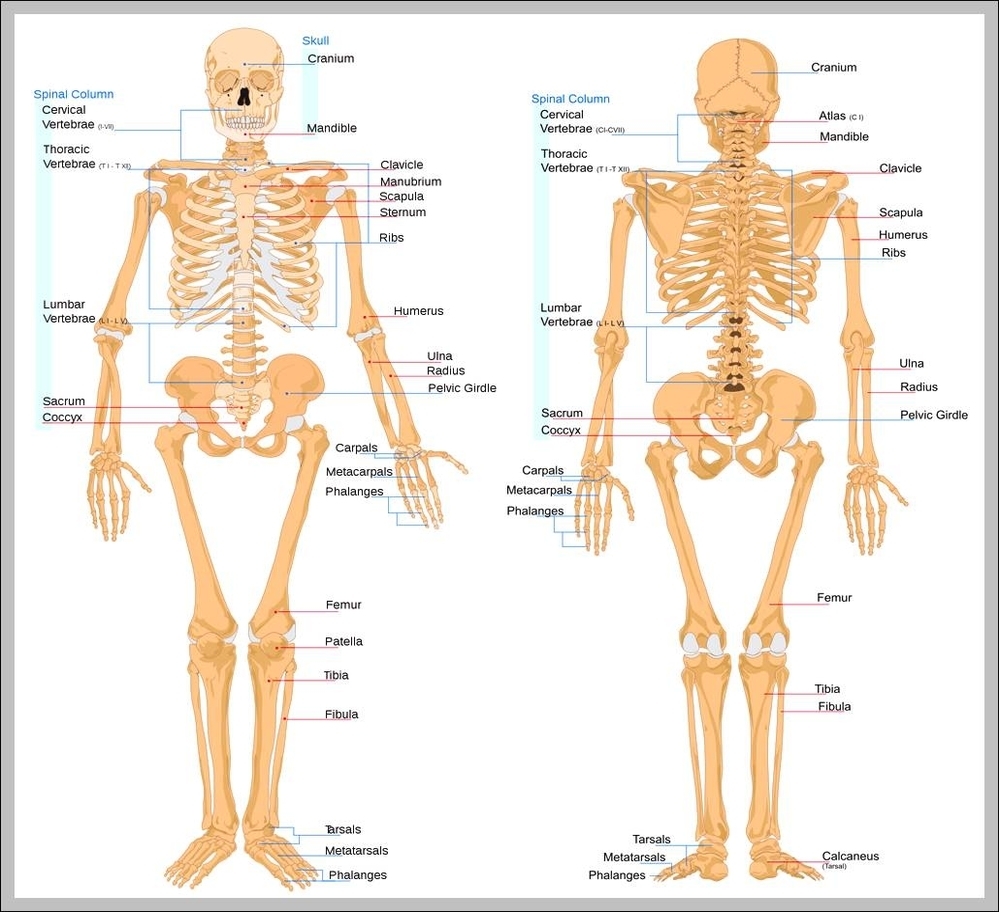
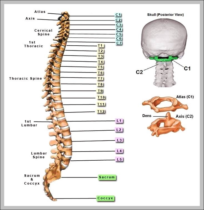
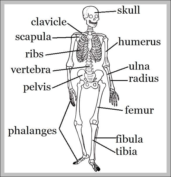
List of bones of the human skeleton. The human skeleton of an adult consists of 206-208 bones. It is composed of 270 bones at birth, but later decreases to 80 bones in the axial skeleton and 126 bones in the View Diagram Pictures Of Bones In The Human Body