Anatomy Of The Lower Leg Achilles Tendon Image

This human anatomy diagram with labels depicts and explains the details and or parts of the Anatomy Of The Lower Leg Achilles Tendon Image. Human anatomy diagrams and charts show internal organs, body systems, cells, conditions, sickness and symptoms information and/or tips to ensure one lives in good health.

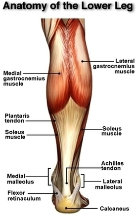
The Achilles tendon is also located in the lower leg. The lower leg is made up of two very strong, long bones: the tibia and the fibula. The tibia, also known as the shin bone, is the stronger and larger of the two. It is located toward the middle of the lower leg. The fibula, or calf bone, is smaller and is located on the outside of the lower leg.

The Achilles tendon attaches the muscles of the calves to the bones of the ankle and foot. The ankle is a joint that connects the lower leg to the foot.

Pain in the back of the heel, worse with shoes on, is the common symptom. Physical examination: To look for Achilles tendon problems, an examiner checks for pain, swelling, warmth, thickening, or discoloration around the heel and leg. A knot on the back of the leg may be present with Achilles tendon rupture.

Anatomy Of The Lower Leg Achilles Tendon Image

Anatomy Of The Lower Leg Achilles Tendon Image


Tags: , , ,