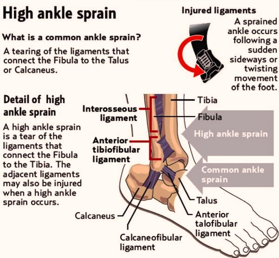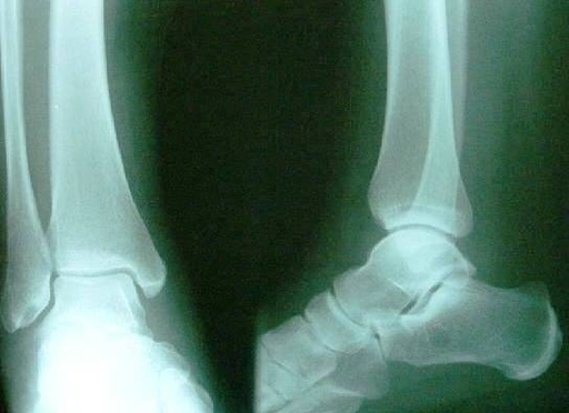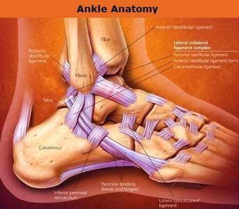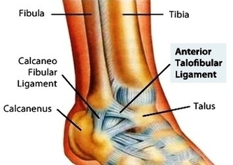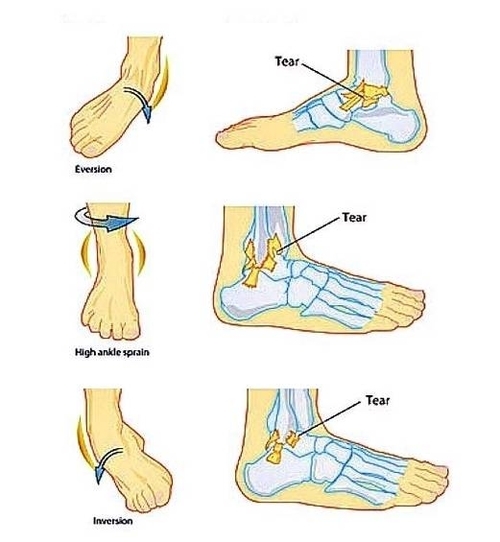
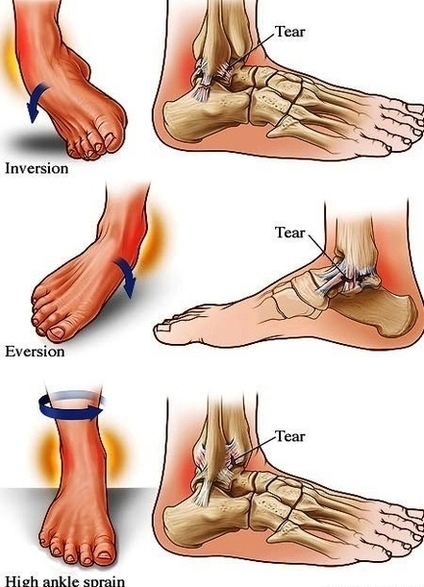
Ankle Sprain Anat1 Image

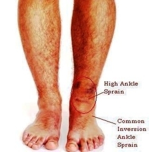
Bruising and swelling are common signs of a sprained ankle. If there is severe tearing of the ligaments, you might also hear or feel a “pop” when the sprain occurs. Most sprained ankles occur in the lateral ligaments on the View Diagram Ankle Sprain Anat1 Image