Ankle Syndesmosis Diagram Image

This human anatomy diagram with labels depicts and explains the details and or parts of the Ankle Syndesmosis Diagram Image. Human anatomy diagrams and charts show internal organs, body systems, cells, conditions, sickness and symptoms information and/or tips to ensure one lives in good health.

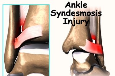
High Ankle Sprain & Syndesmosis Injuries are traumatic injuries that affect the distal tibiofibular ligaments and most commonly occur due to sudden external rotation of the ankle. Diagnosis is suspected clinically with tenderness over the syndesmosis which worsens with squeezing of the tibia and fibula together at the midcalf.

Arthrodesis of the ankle syndesmosis may also be performed as a salvage procedure in patients with pain, arthritis, and chronic syndesmotic instability. This procedure is indicated when other strategies have failed.

The diagnosis of syndesmosis injuries is usually made by examining the ankle. The doctor moves your ankle in different positions in order to check the ligaments and tendons around the ankle. The syndesmosis is stressed by turning the ankle outward while holding the lower leg still.

Ankle Syndesmosis Diagram Image

Ankle Syndesmosis Diagram Image


Tags: ,