Ankle Anatomy Image

This human anatomy diagram with labels depicts and explains the details and or parts of the Ankle Anatomy Image. Human anatomy diagrams and charts show internal organs, body systems, cells, conditions, sickness and symptoms information and/or tips to ensure one lives in good health.

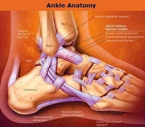
The anatomy of the foot is incredibly complex. This introduction to the anatomy of the foot and ankle will be very general and highlight the most relevant structures. The ankle joint or tibiotalar joint is formed where the top of the talus (the uppermost bone in the foot) and the tibia (shin bone) and fibula meet.

8,460 ankle anatomy stock photos and images available, or search for foot and ankle anatomy to find more great stock photos and pictures. The ankle joint, tendons of the ankle joint foot anatomy vector… bones of the foot and ankle joint medical vector illustration… Houman body parts flat line icons set.

There are many different muscles and ligaments in the ankle, which gives the ankle its strength, flexibility, and range of motion. Save. Anterior and posterior ankle ligaments. Ligaments are a type of soft tissue that is made up mostly of collagen.

Ankle Anatomy Image

Ankle Anatomy Image


Tags: