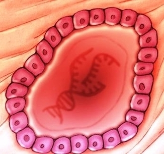
Diagram Of Type Diabetes Metabolic Process Image
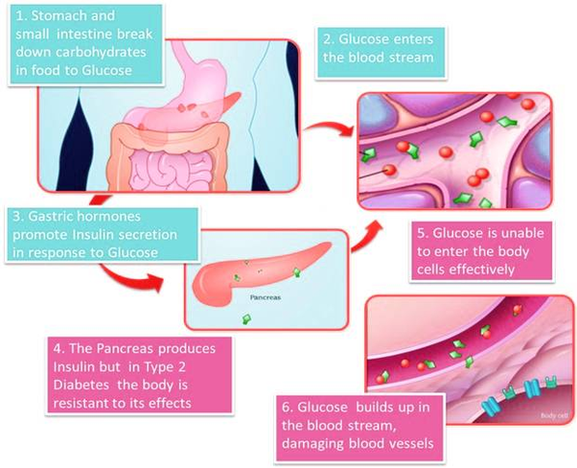
In type 1 diabetes, this happens because the immune system is attacking the cells that make insulin, which are in the pancreas. In type 2 diabetes, the body stops responding to insulin as well as it should. In this article, View Diagram Diagram Of Type Diabetes Metabolic Process Image