Capillary Microcirculation Diagram Image

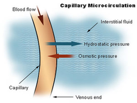
Scott Sundick, MD, is a board-certified vascular and endovascular surgeon. He currently practices in Westfield, New Jersey. Capillaries are the smallest blood vessels in the body, connecting the smallest arteries to the smallest veins. These vessels are often referred to as the “microcirculation.”

Microcirculation Structure and Function. The microcirculation is comprised of arterioles, capillaries, venules, and terminal lymphatic vessels. Small precapillary resistance vessels (10-200 μ) composed of an endothelium surrounded by one or more layers of smooth muscle cells.

They are present in muscle, skin, fat, and nerve tissue. Fenestrated: These capillaries have small pores that allow small molecules through and are located in the intestines, kidneys, and endocrine glands. Sinusoidal or discontinuous: These capillaries have large open pores—large enough to allow a blood cell through.

Capillary Microcirculation Diagram Image Diagram - Chart - diagrams and charts with labels. This diagram depicts Capillary Microcirculation Diagram Image

Capillary Microcirculation Diagram Image

Capillary Diagram Image

27 Capillary Diagram Premium High Res Photos 27 capillary diagramstock photos and images available or start a new search to explore more stock photos and images. venous and arterial – capillary diagram stock pictures, royalty-free photos & images Diagram Demonstrating The Progression Of An Arterial Embolism.

Capillaries are composed of only the tunica intima, consisting of a thin wall of simple squamous endothelial cells. They are the smallest blood vessels in the body: they convey blood between the arterioles and venules.

Simplified image showing blood-flow through the body, passing through capillary networks in its path. The capillary wall performs an important function by allowing nutrients and waste substances to pass across it.

Capillary Diagram Image Diagram - Chart - diagrams and charts with labels. This diagram depicts Capillary Diagram Image

Capillary Diagram Image

Lymph Capillary Diagram Image

Lymph capillaries or lymphatic capillaries are tiny, thin-walled microvessels located in the spaces between cells (except in the central nervous system and non-vascular tissues) which serve to drain and process extracellular fluid. Upon entering the lumen of a lymphatic capillary, the collected fluid is known as lymph.

Lymph capillary. Attached to the lymphatic capillaries are anchoring filaments, which contain elastic fibers. They extend out from the lymphatic capillary, attaching lymphatic endothelial cells to surrounding tissues. When excess interstitial fluid accumulates and causes tissue swelling, the anchoring filaments are pulled,…

Upon entering the lumen of a lymphatic capillary, the collected fluid and associated cells (notably white blood cells) is known as lymph. Each lymphatic capillary carries lymph into a lymphatic vessel, which in turn connects to a lymph node. Lymph is ultimately returned to the venous circulation .

Lymph Capillary Diagram Image Diagram - Chart - diagrams and charts with labels. This diagram depicts Lymph Capillary Diagram Image

Lymph Capillary Diagram Image