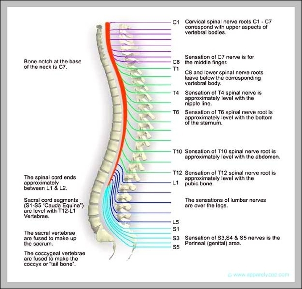
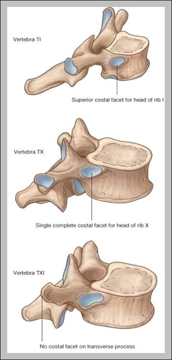
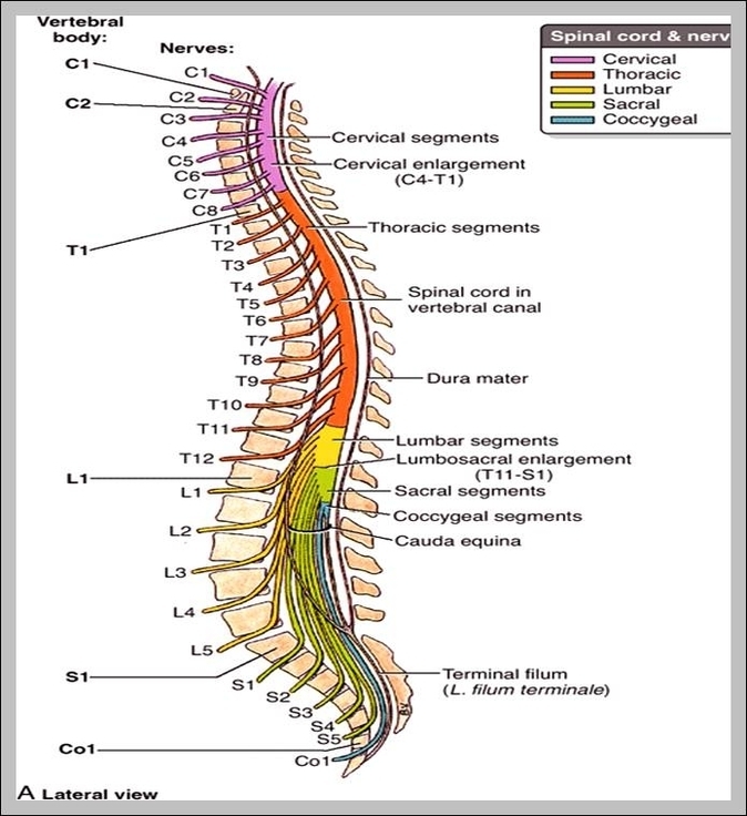
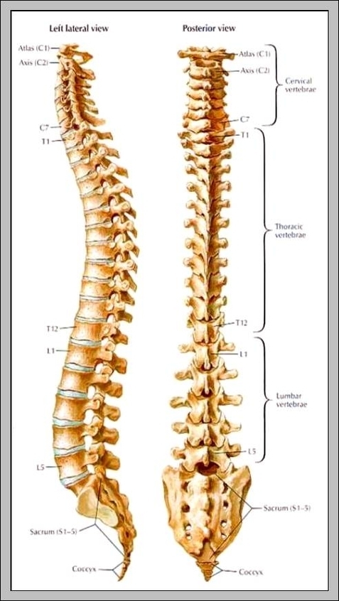
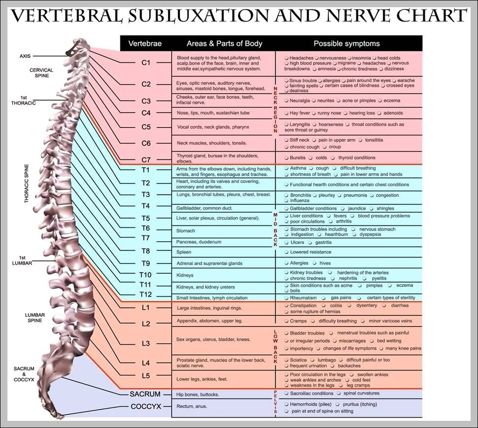
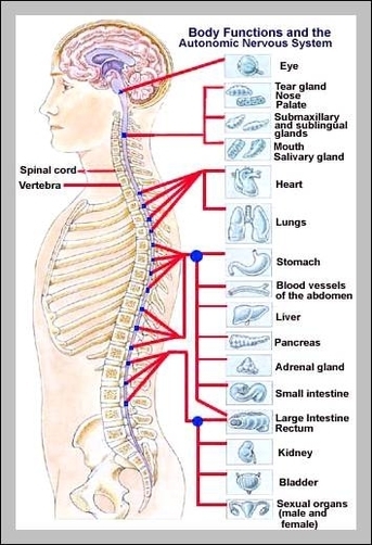
Spinal Cord Vertebrae

Spinal Cord Vertebrae

The spinal cord runs inside the vertebral column, and vertebral column protects the spinal cord. This is the key difference between spinal cord and vertebral column. Spinal cord and vertebral column are two vital structures of humans. They run together View Diagram Spinal Cord Vertebrae

Skeleton Structure

Skeleton Structure

Skeleton defines as the structure of a framework which consists of all bones in the human body or animal. The structure of these bones helps the body of animal s or humans in movement, walking, and performs different other tasks View Diagram Skeleton Structure

Skull Bone Anatomy

Skull Bone Anatomy

Some of the sutures in the skull take a few months-to-years after birth to completely form. The brain is encased in the cranium of the skull. The bones that make up the cranium are called the cranial bones. The remainder View Diagram Skull Bone Anatomy

Picture Of Human Skeleton With Muscles

Picture Of Human Skeleton With Muscles

There are around 640 skeletal muscles within the typical human body. There are around 640 skeletal muscles within the typical human body. Almost every muscle constitutes one part of a pair of identical bilateral muscles, found on both sides, resulting View Diagram Picture Of Human Skeleton With Muscles

Skeletal System Foot

Skeletal System Foot

Medically reviewed by Healthline’s Medical Network on April 9, 2015. The skeletal structure of the foot is similar to that of the hand but, because the foot bears more weight, it is stronger but less movable. The bones of the View Diagram Skeletal System Foot

Skeletal Muscle Picture

Skeletal Muscle Picture

Skeletal muscles are the muscles that we can see and feel through our skin. An individual skeletal muscle is considered an organ within the muscular system of the body. The skeletal muscle works with nerve tissue, connective tissue and vascular View Diagram Skeletal Muscle Picture

Skeleton Bone Names

Skeleton Bone Names

Best Skeleton Names The Big Le-bone-ski Bones McCoy Teddy Bones-evelt Bury White Clarence Marrow Johnny Rotten Jon Bone Jovi Hugh Maris Indiana Bones Blake Skeleton Clay Vackle Albert Spinestein Vincent Van Bone Scary Potter Jim Reaper Eddie Deader Doug Upp View Diagram Skeleton Bone Names

Spinal Vertebrae Chart

Spinal Vertebrae Chart

These regions are called the cervical spine, thoracic spine, lumbar spine, sacrum and coccyx. There are seven cervical vertebrae, twelve thoracic vertebrae and five lumbar vertebrae. The number of vertebrae in a region can vary but overall the number remains View Diagram Spinal Vertebrae Chart

Skeletal Feet

Skeletal Feet

However, many muscles that power the foot’s movement originate as high up as the back of the knee. Some important muscles that affect the foot include: Soleus: This muscle extends from the back of the knee to the heel. It View Diagram Skeletal Feet

Skeletal System Pics

Skeletal System Pics

Teeth are not bones but are still part of the human skeletal system. While the adult human skeletal system includes 206 bones, the infantile skeletal system has many more, since not all of the bones have fused together yet. The View Diagram Skeletal System Pics

Picture Of Skeleton With Names Of Bones

Picture Of Skeleton With Names Of Bones

The main bones of the human skeleton are: The Skull – Cranium, Mandible, and Maxilla. Shoulder girdle – clavicle and scapula – humerus, radius, and ulna. Hand – Carpals, Metacarpals, and Phalanges. Chest – Sternum, and Ribs. Spine – Cervical View Diagram Picture Of Skeleton With Names Of Bones

Picture Of Humerus Bone

Picture Of Humerus Bone

In the upper arm, the brachial artery branches into several arteries, distributing oxygenated blood from the lungs and heart. The radial nerve runs a similar course over the bone and into the forearm. Because it connects at the shoulder with View Diagram Picture Of Humerus Bone

Skeletal Muscle Labeled

Skeletal Muscle Labeled

Skeletal Muscle Labeled: A skeletal muscle labeled diagram highlights the major skeletal muscles in the body, such as the quadriceps, biceps, and deltoids, along with their names and locations.

Hand Wrist Bones

Hand Wrist Bones

Hand Wrist Bones: The wrist is composed of eight carpal bones that connect the hand to the forearm, while the hand includes metacarpals and phalanges, allowing for a wide range of motion and dexterity.

Hand Bones Pictures

Hand Bones Pictures

Hand Bones Pictures: Pictures of hand bones show the skeletal structure of the hand, which consists of 27 bones: 8 carpal bones, 5 metacarpals, and 14 phalanges. These bones allow for dexterity and gripping ability.