Picture Of Skeleton With Names Of Bones

This human anatomy diagram with labels depicts and explains the details and or parts of the Picture Of Skeleton With Names Of Bones. Human anatomy diagrams and charts show internal organs, body systems, cells, conditions, sickness and symptoms information and/or tips to ensure one lives in good health.

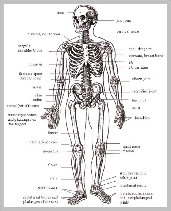
The main bones of the human skeleton are: The Skull – Cranium, Mandible, and Maxilla. Shoulder girdle – clavicle and scapula – humerus, radius, and ulna. Hand – Carpals, Metacarpals, and Phalanges. Chest – Sternum, and Ribs. Spine – Cervical area (top 7 vertebrae), Thoracic (next 12), Lumbar (bottom …

The skeleton can be divided into two parts known as axial and the appendicular. The axial skeleton consists of the central core of the skull, spine, and ribs whilst the appendicular is composed of the arms and legs. How are bones formed? Bones are formed by the ossification of cartilage.

The skeletal system in an adult body is made up of 206 individual bones. These bones are arranged into two major divisions: the axial skeleton and the appendicular skeleton. The axial skeleton runs along the body’s midline axis and is made up of 80 bones in the following regions: Skull.

Picture Of Skeleton With Names Of Bones

picture of skeleton with names of bones


Tags: , , , , , , , , , , , , , , , , , , ,