Skeletal Feet

This human anatomy diagram with labels depicts and explains the details and or parts of the Skeletal Feet. Human anatomy diagrams and charts show internal organs, body systems, cells, conditions, sickness and symptoms information and/or tips to ensure one lives in good health.

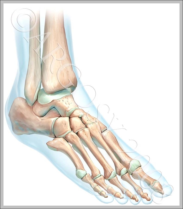
However, many muscles that power the foot’s movement originate as high up as the back of the knee. Some important muscles that affect the foot include: Soleus: This muscle extends from the back of the knee to the heel. It is pivotal in walking and standing.

The feet are divided into three sections: The forefoot contains the five toes (phalanges) and the five longer bones (metatarsals). The midfoot is a pyramid-like collection of bones that form the arches of the feet. These include the three cuneiform bones, the cuboid bone, and the navicular bone.

It extends slightly lower than the medial malleolus forming the outer side of the ankle joint where it articulates with (meets) the talus. The talus is the highest foot bone. It forms the bottom of the ankle joint, articulating with the tibia and fibula (shin bones) and the top of the subtalar joint, articulating with the calcaneus (heel bone).

Skeletal Feet

skeletal feet


Tags: , , , , , , , , , , ,