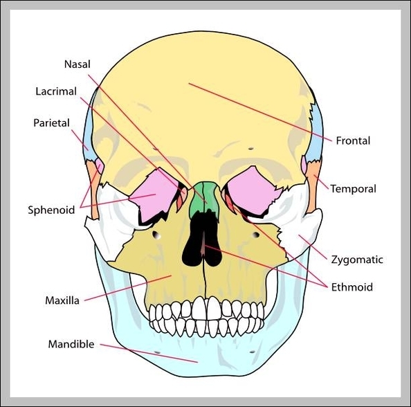
Pictures Of Bones In The Human Body

Pictures Of Bones In The Human Body

List of bones of the human skeleton. The human skeleton of an adult consists of 206-208 bones. It is composed of 270 bones at birth, but later decreases to 80 bones in the axial skeleton and 126 bones in the View Diagram Pictures Of Bones In The Human Body

Skeleton Human

Skeleton Human

Author of Textbook of Comparative Histology. Human skeleton, the internal skeleton that serves as a framework for the body. This framework consists of many individual bones and cartilages. There also are bands of fibrous connective tissue —the ligaments and the View Diagram Skeleton Human

Pictures Of The Bones In The Human Body

Pictures Of The Bones In The Human Body

The bones provide a structural framework and protection to the soft organs. They also provide for the attachment of muscles, and help us move around. The number of bones in the human body at birth is 300. However, as a View Diagram Pictures Of The Bones In The Human Body

What Are The Functions Of The Skeletal System

What Are The Functions Of The Skeletal System

The skeletal system is the body system composed of bones and cartilage and performs the following critical functions for the human body: supports the body. facilitates movement. protects internal organs. produces blood cells. stores and releases minerals and fat. The View Diagram What Are The Functions Of The Skeletal System

Skeleton Chart

Skeleton Chart

The Human Skeletal System Chart shows anterior, lateral and posterior views of the skeletal system. This printout will help you learn about the human skeleton and identify some of the important features of our skeletal anatomy. This document contains an View Diagram Skeleton Chart

Skeleton Labeled Diagram

Skeleton Labeled Diagram

Labeled Human Skeleton. The labeled human skeleton system is comprised of 206 different bones of various sizes and shapes, all with the primary purpose of providing support, protection, and shape to the human body. When humans are born we have View Diagram Skeleton Labeled Diagram

Skeletal Pictures

Skeletal Pictures

Teeth are not bones but are still part of the human skeletal system. While the adult human skeletal system includes 206 bones, the infantile skeletal system has many more, since not all of the bones have fused together yet. The View Diagram Skeletal Pictures

Picture Of Skull Bones

Picture Of Skull Bones

The bones that make up the cranium are called the cranial bones. The remainder of the bones in the skull are the facial bones. Figure 6.7 and Figure 6.8 show all the bones of the skull, as they appear from View Diagram Picture Of Skull Bones

Skeletal Muscle Picture 2

Skeletal Muscle Picture 2

Skeletal muscle, also called voluntary muscle, in vertebrates, most common of the three types of muscle in the body. Skeletal muscles are attached to bones by tendons, and they produce all the movements of body parts in relation to each View Diagram Skeletal Muscle Picture 2

Pictures Of Skeletal System

Pictures Of Skeletal System

Skeletal System 1 Skull. 2 Hyoid and Auditory Ossicles. 3 Vertebrae. 4 Ribs and Sternum. 5 Pectoral Girdle and Upper Limb. 6 Pelvic Girdle and Lower Limb. 7 Microscopic Structure of Bones. 8 Types of Bones. 9 Parts of Bones. View Diagram Pictures Of Skeletal System

Picture Of Human Vertebrae

Picture Of Human Vertebrae

The human spine is composed of 33 vertebrae that interlock with each other to form the spinal column. The human spinal cord consists of nerves that connect the brain to nerves in the body. The vertebrae of the spine align View Diagram Picture Of Human Vertebrae

T5 Vertebrae

T5 Vertebrae

The 12 thoracic vertebrae are labeled T1 through T12, with T1 being closest to the skull and T12 being closest to the tailbone. The T5 is the fifth thoracic vertebra closest to the skull. Most thoracic vertebrae have a variety View Diagram T5 Vertebrae

Thoracic Vertebrae Numbers

Thoracic Vertebrae Numbers

The thoracic spine is comprised of 12 vertebrae labeled T1 through T12. The top thoracic vertebra, T1, connects with C7 in the cervical spine above while the bottom thoracic vertebra, T12, connects with L1 in the lumbar spine below. In View Diagram Thoracic Vertebrae Numbers

T 1 Vertebrae

T 1 Vertebrae

The T1 vertebra is the first (uppermost) of the twelve (12) thoracic vertebrae that make up the central and largest section of the spinal column between the lumbar vertebrae below and the cervical vertebrae above. While larger than the C7 View Diagram T 1 Vertebrae

Skeleton Foot

Skeleton Foot

The skeleton of the foot from behind forward is composed of the following bones: Tarsals. Metatarsals. Phalanges. Identification of bones in the skeleton of the foot: Calcaneus (heel bone) is the largest and most proximal bone. Talus is the 2nd View Diagram Skeleton Foot