Pictures Of Vertebrae

This human anatomy diagram with labels depicts and explains the details and or parts of the Pictures Of Vertebrae. Human anatomy diagrams and charts show internal organs, body systems, cells, conditions, sickness and symptoms information and/or tips to ensure one lives in good health.

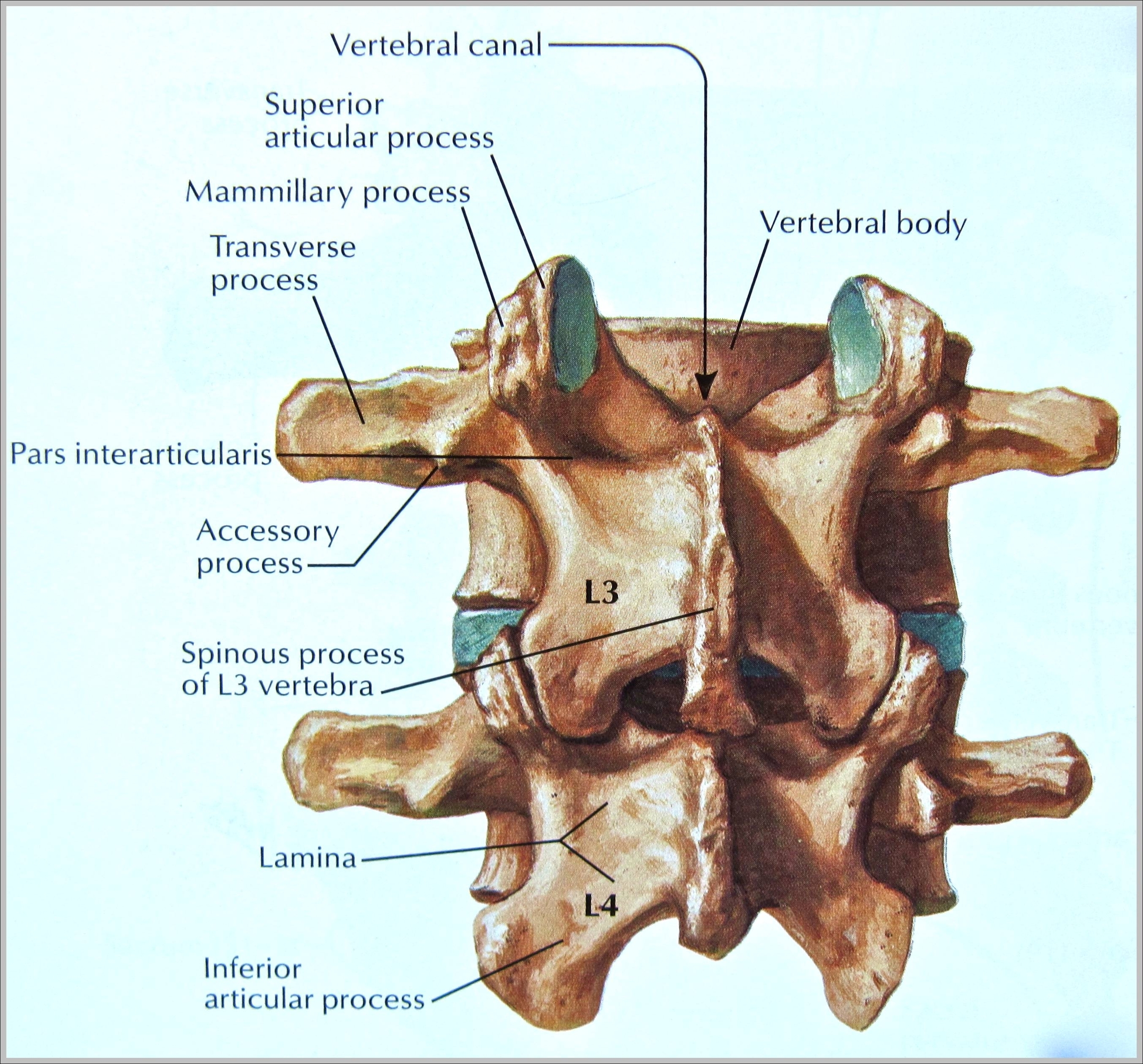
A unique feature of the thoracic vertebrae is that each one forms joints with a pair of ribs to form the sturdy rib cage that protects the organs of the chest. Lumbar: The 5 vertebrae in the lower back form the lumbar region of the spine.

The lumbar vertebrae consist of five individual cylindrical bones that form the spine in the lower back. These vertebrae carry all of the upper body’s weight while providing flexibility and movement to the trunk region.

The vertebrae of the spine align so that their vertebral canals form a hollow, bony tube to protect the spinal cord from external damage and infection. Between the vertebrae are small spaces known as intervertebral canals that allow spinal nerves to exit the spinal cord and connect to the various regions of the body.

Pictures Of Vertebrae

pictures of vertebrae


Tags: , , , , , , ,