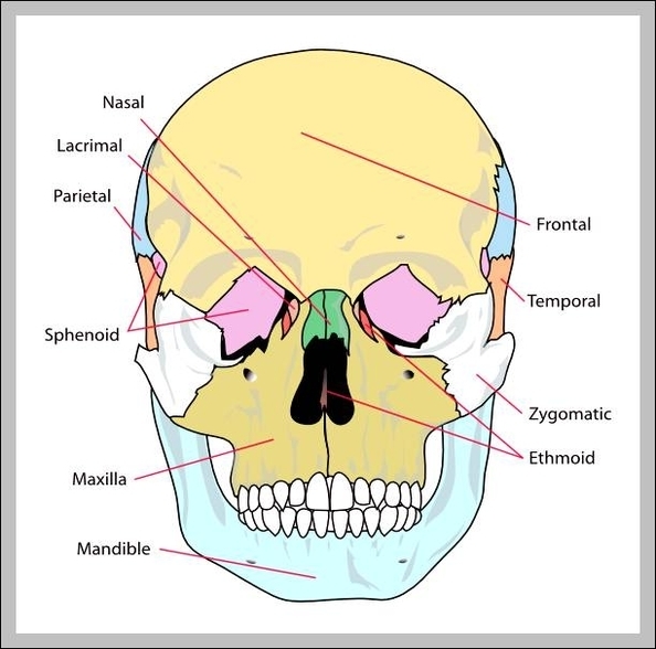
Picture Of Skull Bones

This human anatomy diagram with labels depicts and explains the details and or parts of the Picture Of Skull Bones. Human anatomy diagrams and charts show internal organs, body systems, cells, conditions, sickness and symptoms information and/or tips to ensure one lives in good health.

The bones that make up the cranium are called the cranial bones. The remainder of the bones in the skull are the facial bones. Figure 6.7 and Figure 6.8 show all the bones of the skull, as they appear from the outside. In Figure 6.9, some of the bones of the hard palate forming the roof of the mouth are visible because the mandible is not present.

Occipital bone: the bone that forms the back of the head and connects with the occipital condyles and foramen magnum — skeletal structures located on the underside of the skull, near the spine — and the lambdodial suture, which is at the back of the skull.

A collection of 22 bones, the skull protects the all-important brain and supports the other soft tissues of the head. During fetal development, the bones of the skull form within tough, fibrous membranes in a fetus’ head. As these bones grow throughout fetal and childhood development, they begin to fuse together, forming a single skull.

Picture Of Skull Bones

picture of skull bones


Tags: , , , , , , , , , , ,