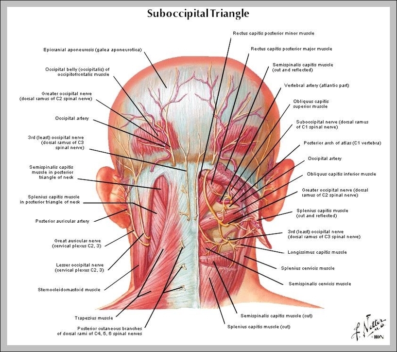
The neck is connected to the upper back through a series of seven vertebral segments. The cervical spine has 7 stacked bones called vertebrae, labeled C1 through C7. The top of the cervical spine connects to the skull, and the bottom connects to the upper back at about shoulder level.
· Longus Colli- Begins between the third and sixth cervical vertebrae, responsible for flexion of the head and neck. · Longus Capitis- Begins between the third and sixth cervical vertebrae, responsible for flexion of the neck · Rectus Capitis Anterior- Begins at the first cervical vertebrae, responsible for flexion of the neck
Truncal vertebrae (divided into thoracic and lumbar vertebrae in mammals) lie caudal (toward the tail) of cervical vertebrae. In sauropsid species, the cervical vertebrae bear cervical ribs. In lizards and saurischian dinosaurs, the cervical ribs are large; in birds, they are small and completely fused to the vertebrae.
Vertebrae In The Neck

