Sexual Differentation
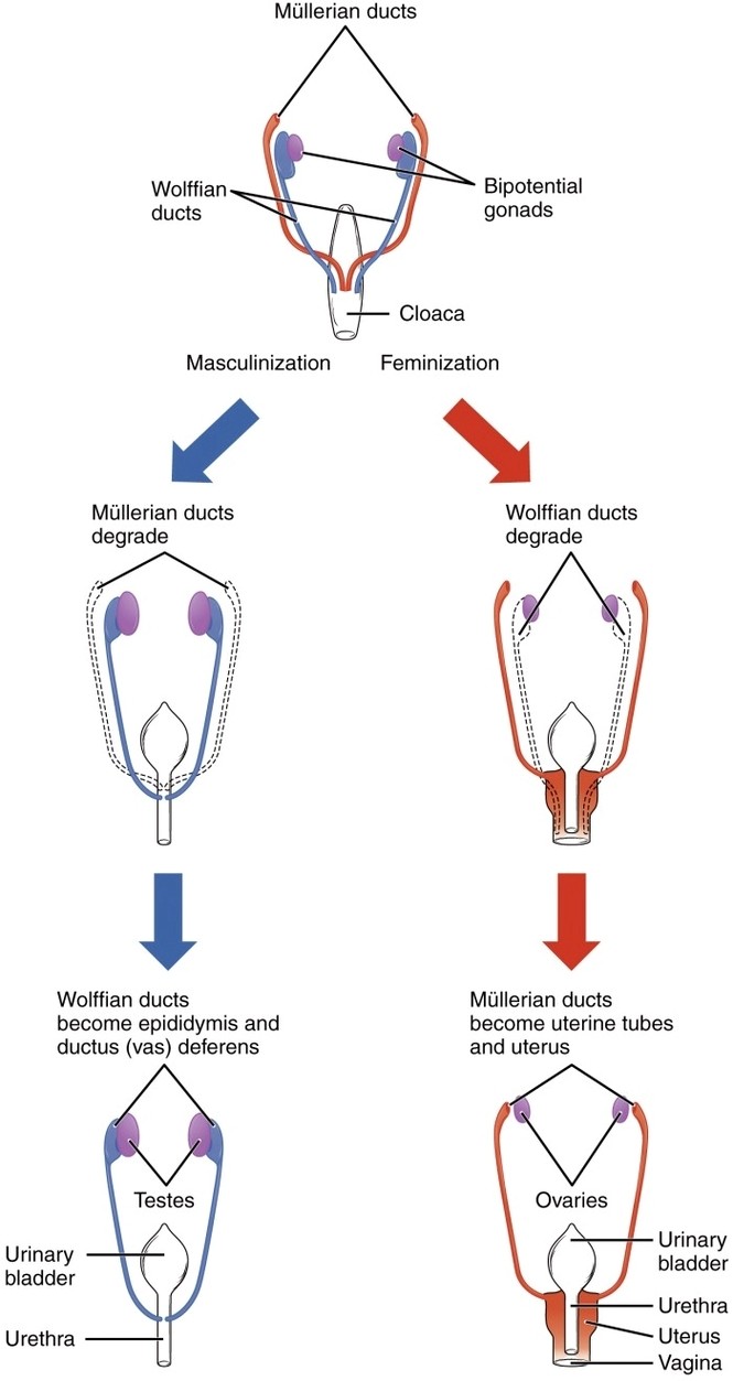
Sexual Differentiation: Sexual differentiation begins with genetic sex (XX or XY), then proceeds under hormonal influence, with testes producing testosterone that drives male development and its absence allowing female structures to develop.