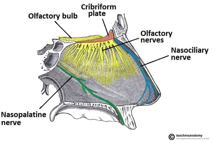
Nasopalatine Nerve and Incisive Canal
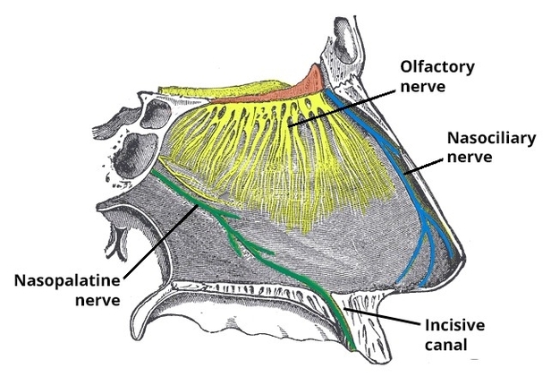
The nasopalatine nerve passes through the incisive canal, providing sensory innervation to the anterior palate and adjacent gingiva. Knowledge of its anatomy is essential for dentists, oral surgeons, and medical students in anesthesia, implant placement, or maxillofacial surgery. Understanding the View Diagram Nasopalatine Nerve and Incisive Canal