Skip to content

Human Anatomy Diagrams

Menu

Primary menu

  • Top
  • Anatomy
  • Muscles
  • Systems
  • Body Parts
  • Organs
  • Cells
  • Bones
  • Teeth
  • Cancer
  • Health

Author Archives: anatomy

Erythroblastosis Fetalis

Posted on July 17, 2025 by anatomy
Erythroblastosis Fetalis

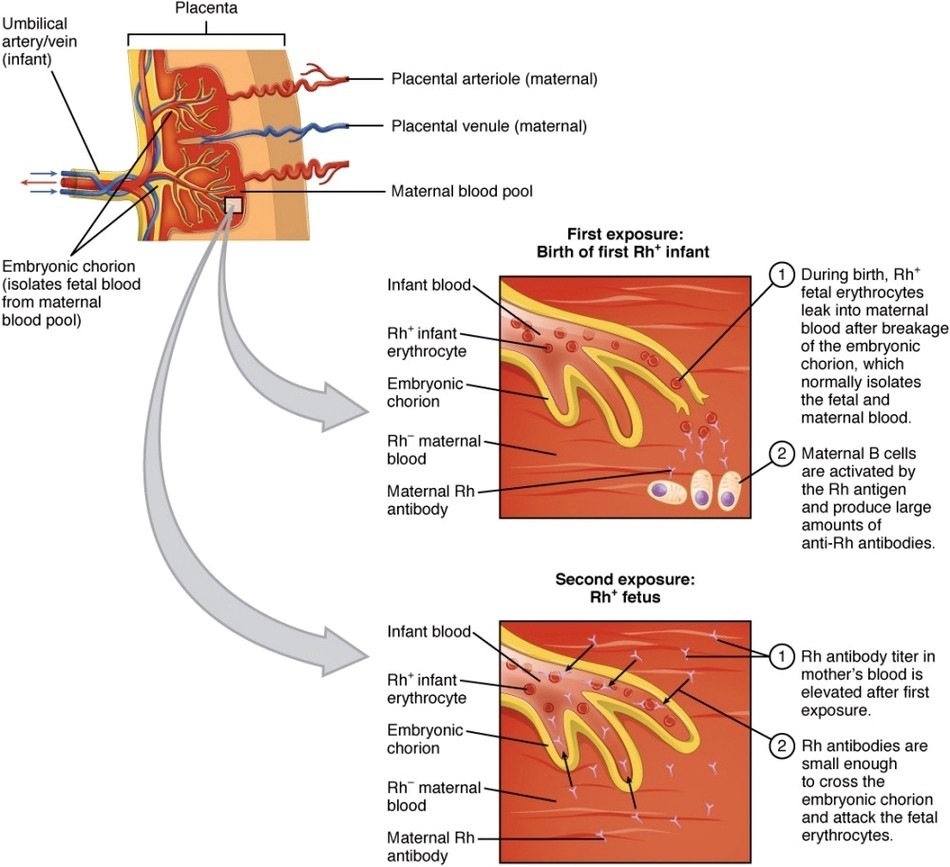
Erythroblastosis Fetalis: Erythroblastosis fetalis is a hemolytic disease of the newborn caused by Rh incompatibility, where maternal antibodies attack fetal red blood cells, leading to anemia and jaundice.

Posted in Cells | Tagged blood disorder, erythroblastosis fetalis diagram, fetal anemia, pregnancy complications, Rh incompatibility

The Posterior Pituitary Complex

Posted on July 16, 2025 by anatomy
The Posterior Pituitary Complex

The Posterior Pituitary Complex: The posterior pituitary stores and releases oxytocin and antidiuretic hormone (ADH), both produced in the hypothalamus, affecting uterine contractions and water reabsorption in the kidneys, respectively.

Posted in Organs | Tagged ADH and oxytocin, endocrine system, hormone release, neurohypophysis function, posterior pituitary diagram

Varicose Vein

Posted on July 15, 2025 by anatomy
Varicose Vein

Varicose Vein: Varicose veins occur when valves in superficial veins become incompetent, causing blood to pool and the veins to become enlarged, twisted, and visible beneath the skin, typically in the legs.

Posted in Body Parts | Tagged blood flow disorders, leg vein anatomy, varicose vein diagram, vascular health, venous insufficiency

Mendelian Pea Plant Cross

Posted on July 14, 2025 by anatomy
Mendelian Pea Plant Cross

Mendelian Pea Plant Cross: Mendel’s pea plant crosses revealed predictable inheritance patterns through dominant and recessive traits, forming the foundation of classical genetics.

Posted in Cells | Tagged dominant recessive traits, genetic experiments, Mendelian inheritance diagram, pea plant cross, Punnett square

Cardiac Conduction

Posted on July 13, 2025 by anatomy
Cardiac Conduction

Cardiac Conduction: The heart’s electrical system begins with the sinoatrial (SA) node, spreading impulses through the atria, to the atrioventricular (AV) node, and down the bundle branches and Purkinje fibers to coordinate contraction.

Posted in System | Tagged AV node, cardiac conduction system diagram, heart electrical pathway, Purkinje fibers, SA node

Lower Limb Arteries Anterior Posterior

Posted on July 12, 2025 by anatomy
Lower Limb Arteries Anterior Posterior

Lower Limb Arteries Anterior Posterior: The anterior and posterior tibial arteries, along with the femoral and popliteal arteries, supply oxygenated blood to the muscles and skin of the leg and foot.

Posted in Anatomy | Tagged arterial circulation, blood supply to legs, femoral artery, lower limb arteries diagram, vascular anatomy

The Location Structure and Histology of the Thymus

Posted on July 11, 2025 by anatomy
The Location Structure and Histology of the Thymus

The Location Structure and Histology of the Thymus: The thymus is located in the anterior mediastinum, made of lobules containing cortex and medulla, and supports T cell maturation, especially in childhood.

Posted in Organs | Tagged immune system, T cell maturation, thymus diagram, thymus histology, thymus structure

Ventricular Muscle Thickness

Posted on July 10, 2025 by anatomy
Ventricular Muscle Thickness

Ventricular Muscle Thickness: The walls of the left ventricle are thicker than the right due to the higher pressure needed to pump blood throughout the body, compared to the lower resistance in the pulmonary circulation.

Posted in Muscles | Tagged cardiac hypertrophy, heart muscle diagram, left vs right ventricle, myocardial anatomy, ventricular wall thickness

Gallbladder

Posted on July 9, 2025 by anatomy
Gallbladder

Gallbladder: The gallbladder stores and concentrates bile produced by the liver, releasing it into the small intestine to aid in the digestion and absorption of fats.

Posted in Organs | Tagged bile storage, digestive system, gallbladder anatomy, gallbladder diagram, liver function

Muscular and Elastic Artery Arteriole

Posted on July 8, 2025 by anatomy
Muscular and Elastic Artery Arteriole

Muscular and Elastic Artery Arteriole: Elastic arteries (like the aorta) buffer pressure from the heart, muscular arteries distribute blood to organs, and arterioles regulate blood flow into capillary beds via vasoconstriction and vasodilation.

Posted in Anatomy | Tagged arteriole structure, blood flow regulation, elastic artery function, muscular artery diagram, vascular system

Complement Cascade and Function

Posted on July 7, 2025 by anatomy
Complement Cascade and Function

Complement Cascade and Function: The complement system is a group of plasma proteins that, once activated, enhance immune responses by promoting inflammation, opsonizing pathogens for phagocytosis, and directly lysing microbial cells through the membrane attack complex.

Posted in Cells | Tagged complement system diagram, immune response, inflammation, pathogen elimination, protein activation

Bone Marrow

Posted on July 6, 2025 by anatomy
Bone Marrow

Bone Marrow: Bone marrow is the primary site of blood cell production, including red and white blood cells and platelets, and contains stem cells that support hematopoiesis.

Posted in Organs | Tagged blood cell production, bone marrow diagram, bone marrow function, hematopoiesis, hematopoietic system

Arteries of the Brain

Posted on July 5, 2025 by anatomy
Arteries of the Brain

Arteries of the Brain: The brain is supplied by the internal carotid and vertebral arteries, which form the Circle of Willis, ensuring consistent cerebral blood flow.

Posted in Anatomy | Tagged blood supply to brain, brain arteries diagram, brain blood flow, cerebral circulation, vascular brain anatomy

Fenestrated Capillary

Posted on July 4, 2025 by anatomy
Fenestrated Capillary

Fenestrated Capillary: Fenestrated capillaries have pores in their walls that allow for rapid exchange of water and small solutes, commonly found in kidneys, intestines, and endocrine glands.

Posted in Anatomy | Tagged blood filtration, capillary function, capillary structure, fenestrated capillary diagram, vascular anatomy

Hyperbaric Chamber

Posted on July 3, 2025 by anatomy
Hyperbaric Chamber

Hyperbaric Chamber: A hyperbaric chamber delivers 100% oxygen at elevated atmospheric pressure, enhancing oxygen delivery to tissues and used therapeutically for conditions such as decompression sickness, carbon monoxide poisoning, and chronic wounds.

Posted in Health | Tagged hyperbaric chamber diagram, medical treatment, oxygen therapy, pressure chamber, wound healing

Histology Small Intestines

Posted on July 2, 2025 by anatomy
Histology Small Intestines

Histology Small Intestines: The small intestine’s histology features villi and microvilli on its mucosal surface, which greatly increase the surface area for nutrient absorption; it also contains glands and lymphatic structures for digestion and immune defense.

Posted in Anatomy | Tagged enterocyte function, intestinal glands, nutrient absorption, small intestine histology diagram, villi structure

Urea Cycle

Posted on July 1, 2025 by anatomy
Urea Cycle

Urea Cycle: The urea cycle occurs in the liver and converts toxic ammonia, a byproduct of protein metabolism, into urea, which is then safely excreted in the urine by the kidneys.

Posted in Cells | Tagged ammonia conversion, liver detoxification, metabolic pathways, nitrogen metabolism, urea cycle diagram

Fetal Circulation

Posted on June 30, 2025 by anatomy
Fetal Circulation

Fetal Circulation: Fetal circulation features shunts like the ductus venosus, foramen ovale, and ductus arteriosus that divert blood away from the liver and lungs, directing it efficiently between the heart and placenta.

Posted in Anatomy | Tagged blood flow during pregnancy, embryonic heart, fetal circulation diagram, fetal development, placenta

Neonatal Circulatory System

Posted on June 29, 2025 by anatomy
Neonatal Circulatory System

Neonatal Circulatory System: At birth, the neonatal circulatory system undergoes major changes as fetal shunts close and the lungs take over oxygen exchange, resulting in the adult-like pattern of blood flow.

Posted in System | Tagged ductus arteriosus, fetal to neonatal transition, foramen ovale closure, infant heart, newborn circulation diagram

The Breakdown of Fatty Acids

Posted on June 28, 2025 by anatomy
The Breakdown of Fatty Acids

The Breakdown of Fatty Acids: Fatty acids are broken down in the mitochondria through a process called beta-oxidation, generating acetyl-CoA, which enters the citric acid cycle to produce ATP for cellular energy.

Posted in Cells | Tagged beta-oxidation, energy production, fatty acid metabolism diagram, lipid catabolism, mitochondrial function

Post navigation

← Older posts
Newer posts →

Primary Sidebar Widget Area

Top Diagrams

  • Human Body Organ Locations Human Body Organ Locations

  • Map Of Body Muscles Map Of Body Muscles

  • Human Organs Back View Human Organs Back View

  • Spinal Vertebrae Chart Spinal Vertebrae Chart

  • Female Abdominal Anatomy Female Abdominal Anatomy

  • Map Of Human Organs Map Of Human Organs

  • Male Body Organs Diagram Male Body Organs Diagram

  • Female Muscular System Female Muscular System

  • Lower Back Hip Anatomy Lower Back Hip Anatomy

  • Picture Of Organs On Left Side Of Body Picture Of Organs On Left Side Of Body

  • Male Anatomy Pictures Male Anatomy Pictures

  • Lower Back Anatomy Pictures Lower Back Anatomy Pictures

  • Female Human Anatomy Organs Female Human Anatomy Organs

  • Muscular System Full Body Diagram Muscular System Full Body Diagram

  • Groin Muscles Anatomy Groin Muscles Anatomy

Latest Posts

  • Aortic Aneurysm
  • Great Vessels of the Heart
  • Inferior Alveolar Nerve Course Supply
  • Sagittal View of the Subdivisions of the Peritoneum
  • Ligaments of the Elbow Joint Lateral and Medial Aspect
  • Borders of the Femoral Triangle
  • Palmar Fascia of the Hand
  • Knee 3D
  • Anatomical Position of the Lungs
  • X Ray of Shoulder Separation
  • Aberrant and Supernumerary Arteries of the Kidney
  • Anatomical Location of the Lacrimal Gland
  • Superficial Cerebral Veins
  • Overview of the Anatomical Position of the Ulna
  • Human Skeletal System Diagram

Categories

  • Anatomy
  • Body Parts
  • Bones
  • Cancer
  • Cells
  • Health
  • Muscles
  • Organs
  • System
  • Teeth
  • Top

Top Tags

  • muscles (205)
  • muscle (171)
  • human (156)
  • human muscles (138)
  • muscles diagram (138)
  • muscles anatomy (138)
  • human muscle diagram (136)
  • muscular system (136)
  • body (119)
  • system (119)
  • body organs (119)
  • blood (82)
  • parts of a human body (77)
  • human body parts (77)
  • body parts (77)
  • organs (67)
  • organ (67)
  • pregnancy (66)
  • diabetes (62)
  • organ anatomy (62)
Copyright © 2026 . All Rights Reserved.
Theme: Catch Box by Catch Themes
Scroll Up