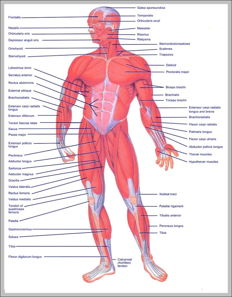
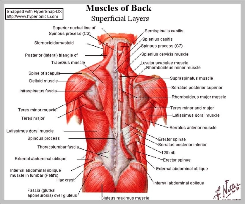
Upper Body Muscle Names
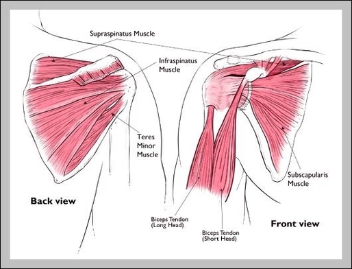
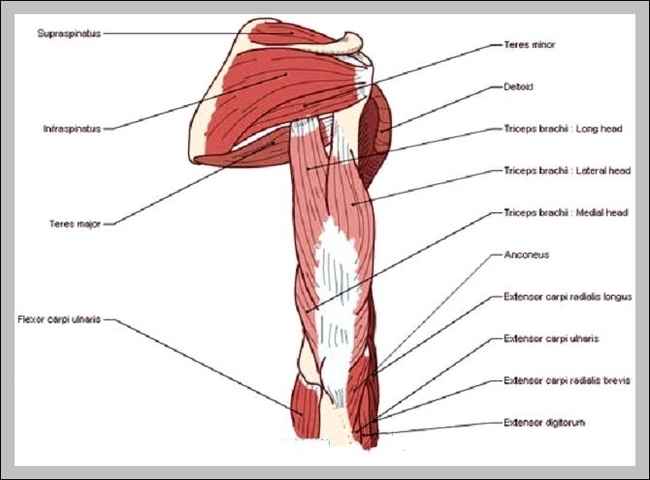
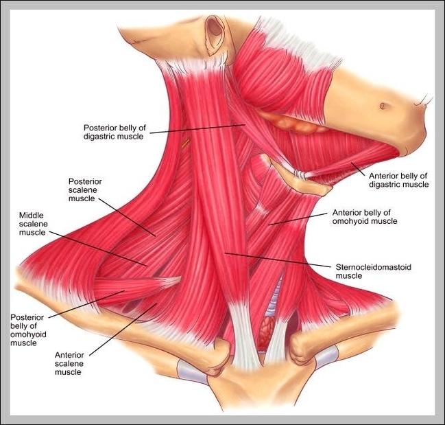
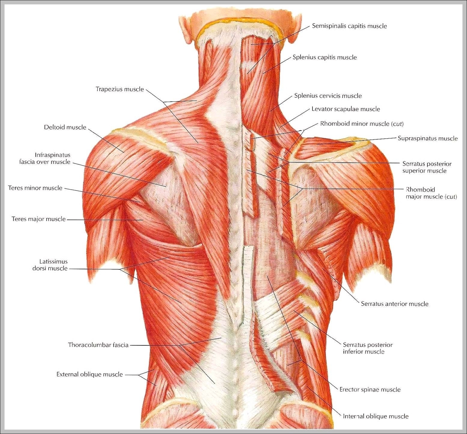
List of Upper Body Muscles Neck and Shoulders. The neck primarily consists of the sternocleidomastoid and the splenius muscles,… Chest. The chest region includes two muscle groups and one individual muscle. Abdominals. The abdominals primarily consist of the rectus abdominis, View Diagram Upper Body Muscle Names