Colon Cancer Stage Image

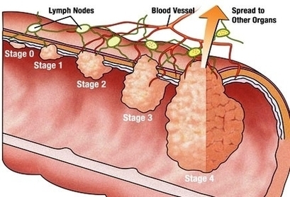
At its earliest stage (stage 0), colon cancer is limited to the inner lining of your colon. As colon cancer progresses, it can grow through your colon and extend to nearby structures. The most advanced stage of colon cancer (stage IV) indicates cancer has spread to other areas of the body, such as the liver or lungs.

It helps determine how serious the cancer is and how best to treat it. Doctors also use a cancer’s stage when talking about survival statistics. The earliest stage colorectal cancers are called stage 0 (a very early cancer), and then range from stages I (1) through IV (4). As a rule, the lower the number, the less the cancer has spread.

After someone is diagnosed with colorectal doctors will try to figure out if it has spread, and if so, how far. This process is called staging. The stage of a cancer describes how much cancer is in the body.

Colon Cancer Stage Image Diagram - Chart - diagrams and charts with labels. This diagram depicts Colon Cancer Stage Image

Colon Cancer Stage Image

Diagram Of Stage Ii Prostate Cancer Image

What to Expect from Stage 2 Prostate Cancer 1 Radical prostatectomy. Radical prostatectomy is surgical removal of the prostate. 2 Radiation therapy. Radiation therapy is used to destroy cancer cells. 3 Hormone therapy. Hormone therapy is used to lower male hormone levels or block them…

The main stages of prostate cancer range from I (1) through IV (4). Some stages are split further (A, B, etc). As a rule, the lower the number, the less the cancer has spread. A higher number, such as stage IV, means cancer has spread more. And within a stage, an earlier letter means a lower stage.

This process is called staging. The stage of a prostate cancer describes how much cancer is in the body. It helps determine how serious the cancer is and how best to treat it. Doctors also use a cancer’s stage when talking about survival statistics.

Diagram Of Stage Ii Prostate Cancer Image Diagram - Chart - diagrams and charts with labels. This diagram depicts Diagram Of Stage Ii Prostate Cancer Image

Diagram Of Stage Ii Prostate Cancer Image

Diagram Of Parkdal Stage Wh Image

When starting work on a project, directors and actors will commonly work from a ground plan, which is a theatre stage diagram onto which will be mapped the elements of a particular theatre set. Ground plans help everyone involved keep track of where set pieces and props need to be at any given time.

The parts of a stage that are visible to an audience are generally called the ”proscenium,” though this term is more broadly applicable in box set theatres. If audience members can see an actor, a prop, or a set piece, for instance, then those people and objects are located in the proscenium.

This area is called the apron. The pit is traditionally used by the orchestra in pieces where actors are accompanied by live music. The pit, also called the orchestra pit, is located directly in front of the stage, between the apron and the first row of seats, and is usually lower than floor level to hide the orchestra.

Diagram Of Parkdal Stage Wh Image Diagram - Chart - diagrams and charts with labels. This diagram depicts Diagram Of Parkdal Stage Wh Image

Diagram Of Parkdal Stage Wh Image

Stage Of Third Month Weeks Of Pregnancy Image

This photo shows a baby at 36 weeks of gestational age during the third trimester. During this stage: The waxy coating that protects the skin (vernix) becomes thicker.

A typical pregnancy lasts 40 weeks from the first day of your last menstrual period (LMP) to the birth of the baby. It is divided into three stages, called trimesters: first trimester, second trimester, and third trimester. The fetus undergoes many changes throughout maturation.

After the third month of pregnancy, the embryonic stage is over and the fetal stage begins, so the embryo is now called fetus. The baby-to-be already has all the organs but they are still developing.

Stage Of Third Month Weeks Of Pregnancy Image Diagram - Chart - diagrams and charts with labels. This diagram depicts Stage Of Third Month Weeks Of Pregnancy Image

Stage Of Third Month Weeks Of Pregnancy Image

Diagram Model Embryo To Cell Stage Icon Image

The image of the embryo or the egg in the mother’s womb has a reddish tint. human embryo development stock pictures, royalty-free photos & images The image of the embryo or the egg in the mother’s womb has a… Stages of embryo development Stages of embryo development. Vector flat infographic icons. human embryo development stock illustrations

Mosaic image composed of 9 sections. Also known as a blastocyst where the inner cell mass or embryoblast forms the embryo. The outer layer of cells are the trophoblast. human embryo development stock pictures, royalty-free photos & images Fertilized, high quality, human egg after five days just prior to transfer into the woman’s uterus.

1,727 human embryo development stock photos and images available, or search for ivf or ivf process to find more great stock photos and pictures. Vector human embryo development circle flat icon Vector human embryo development circle with female uterus icon.

Diagram Model Embryo To Cell Stage Icon Image Diagram - Chart - diagrams and charts with labels. This diagram depicts Diagram Model Embryo To Cell Stage Icon Image

Diagram Model Embryo To Cell Stage Icon Image

Stage Vertebra And Spinal Cord Photo Image

2,848 spinal cord diagram stock photos and images available, or search for nervous system or spine illustration to find more great stock photos and pictures. Numbering order of the vertebrae of the human spinal column. Vertebral column: cervical, thoracic and lumbar spine, sacrum and coccyx.

Spine Diagram A diagram of a human female spine showing a side view of the vertebra of the spinal cord within the the body. This is an editable EPS 10 vector illustration with CMYK color space. spinal cord diagram stock illustrations A diagram of a human female spine showing a side view of the vertebra of the spinal cord within the the body.

Outline: Information and pictures of the spine and spinal cord showing C1 to S5 vertebra and which vertebra effect various body functions. The human spinal cord is about 45 cm (18 in) in men and 43 cm (17 in) long in women. The human spine is composed of 33 vertebrae that interlock with each other to form the spinal column.

Stage Vertebra And Spinal Cord Photo Image Diagram - Chart - diagrams and charts with labels. This diagram depicts Stage Vertebra And Spinal Cord Photo Image

Stage Vertebra And Spinal Cord Photo Image