Nose Nasal Cavities Diagram Image

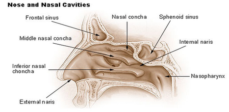
38 nasal cavity diagram stock photos and images available or start a new search to explore more stock photos and images.

White background. nasal cavity stock pictures, royalty-free photos & images The image depicts a near median section through the nose and nasal passages. Details include nasal cavity, soft and hard palate, uvula, eustachian tube and pharyngeal tonsil. White background. Nose anatomy medical vector illustration diagram.

Nasal Cavity Definition. The nose is one of the primary sensory organs responsible for the sense of smell, while it also plays major roles in respiration and speech production [1].

Nose Nasal Cavities Diagram Image Diagram - Chart - diagrams and charts with labels. This diagram depicts Nose Nasal Cavities Diagram Image

Nose Nasal Cavities Diagram Image

Te Nose Diagram Image

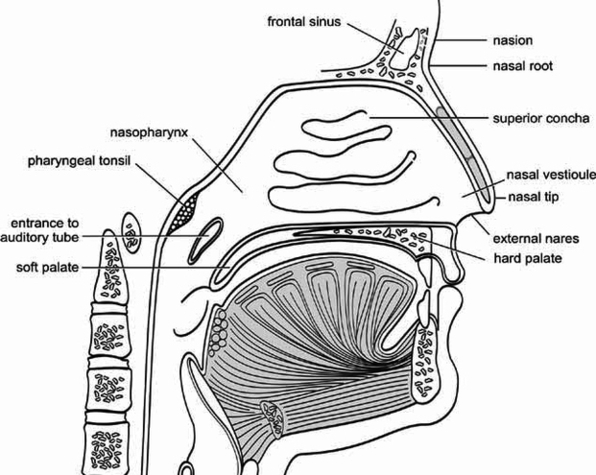
External Structure The surface of the human nose consists of a frontal portion comprised of the glabella, nasion, alar sidewalls and tip points; a basal portion made up of the columella, nostrils, soft tissues and infra tip lobule; and two other portions called the latter and oblique sections.

Determinants for the shape of the nose. Near the tip of the nose are the nostrils and the columella – the strip of skin between the nostrils. The portion of the nostrils that attach to the cheeks is called the alae. Your nose also has four types of muscles called elevators, depressors, compressors and dilators.

The lateral nasal walls, or sidewalls, each contain three finger-like structures called turbinates. These are made of bony core covered with soft tissue and mucosa. The turbinates regulate nasal airflow and provide mucosa surface area.

Te Nose Diagram Image Diagram - Chart - diagrams and charts with labels. This diagram depicts Te Nose Diagram Image

Te Nose Diagram Image