
Tag Archives: nerves
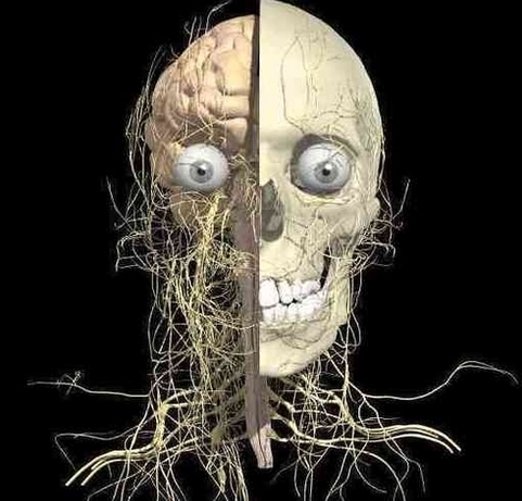
Branial Nerves Brain Image
Cranial Nerves Anatomy Brainstem Human Body En Medical Image
Cranial Nerves Anatomy Brainstem Human Body En Large Photo Image

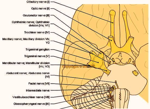
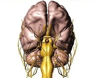
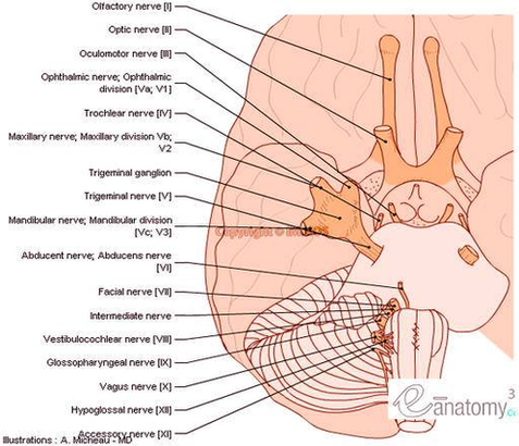
Head Nerves Anatomy Image
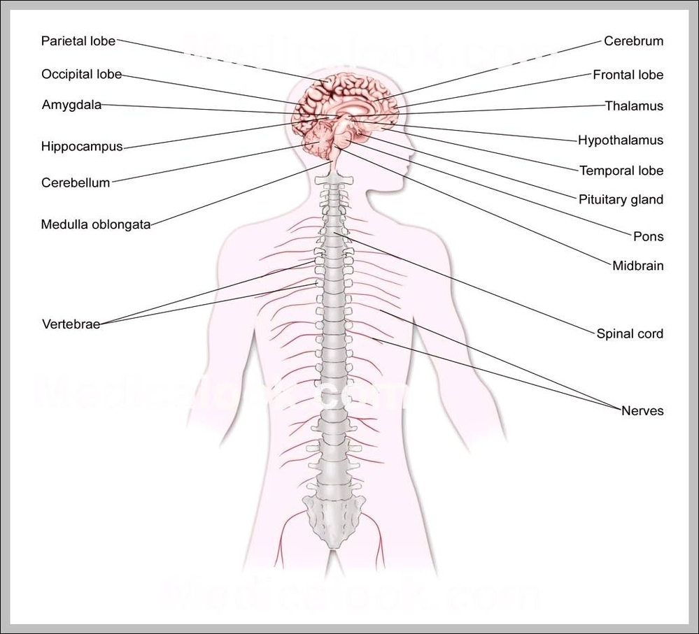
Nerves of the Head and Neck. The nerves of the head and neck include the most vital and important organs of the nervous system — the brain and spinal cord — as well as the organs of the special senses. View Diagram Head Nerves Anatomy Image
Nerves In Back Image
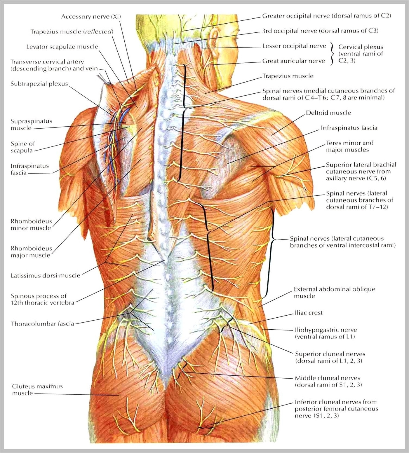
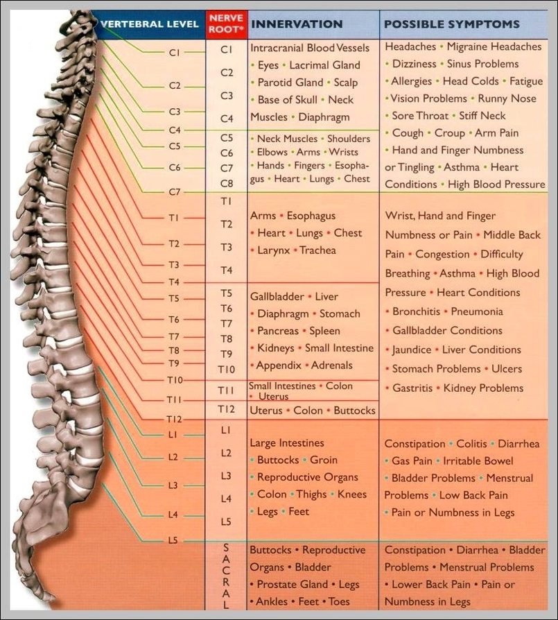
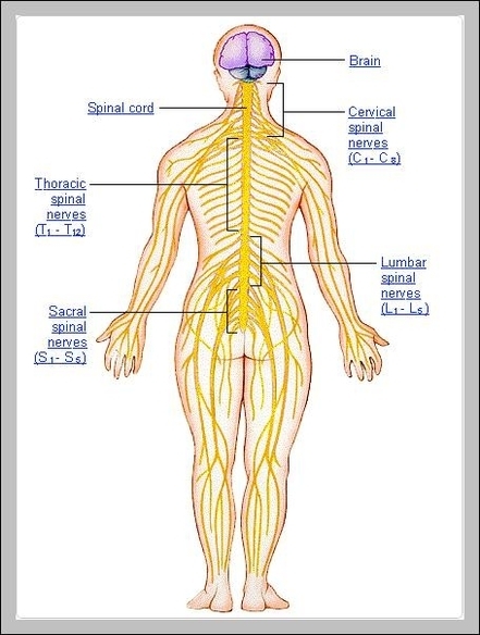
The spinal nerves of the lower back also carry many neurons of the autonomic nervous system (ANS) that maintain the vital involuntary processes of the digestive, urinary, endocrine, and reproductive systems. Spinal Nerves. Here is more information about your nerves, View Diagram Nerves In Back Image
Hand Anatomy Nerves Image
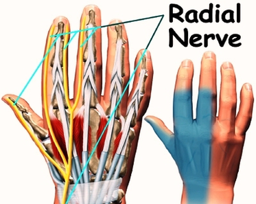
7,371 hand nerve anatomy stock photos, vectors, and illustrations are available royalty-free. See hand nerve anatomy stock video clips The nerve powers almost all of the small muscles in the hand including the hypothenar muscles, the lumbricals to the ring View Diagram Hand Anatomy Nerves Image
Human Body Nerves Image

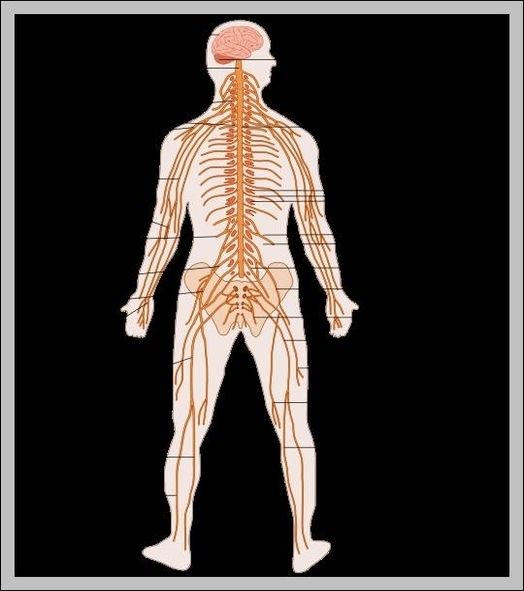
Nerves of the body are a part of a very complicated organ system of the human body, known as the nervous system. They basically form a network of signal carriers, that carry signals to and from the brain to various View Diagram Human Body Nerves Image
Anatomy Arm Nerves Image
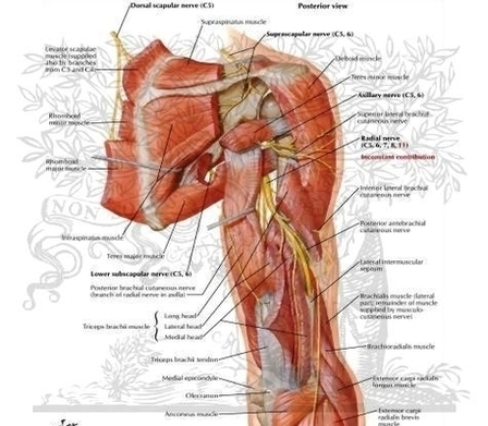
There are four main nerves of the arm and they wrap around the bones. The ulnar and median nerves originate in the brachial plexus, which is a bundle of nerve fibers extending from the spinal cord to the arm and View Diagram Anatomy Arm Nerves Image
What Is The Nervous System
The nervous system is a complex network of nerves and cells that carry messages to and from the brain and spinal cord to various parts of the body. © VectorMine / Shutterstock.com The nervous system is a complex network of View Diagram What Is The Nervous System
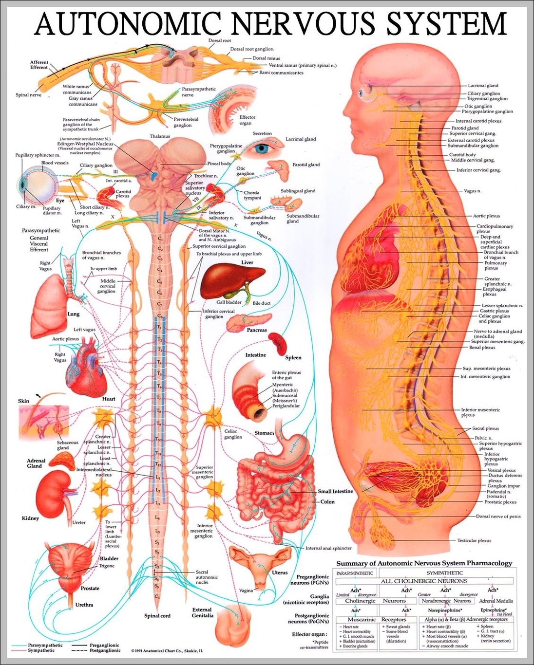
Spinal Nervous System
(The spinal nerves and the area that they innervate are described in the section The peripheral nervous system: Spinal nerves.) central nervous system: humanThe brain and the spinal cord constitute the central nervous system.Created and produced by QA International. Nerves View Diagram Spinal Nervous System
Picture Of Spine And Nerves
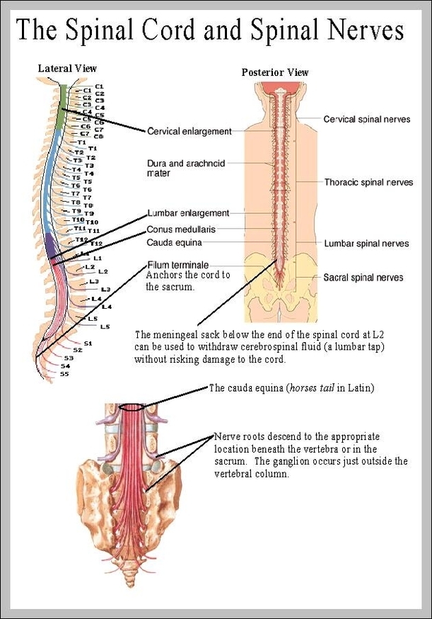
Spinal nerves are the major nerves of the body. A total of 31 pairs of spinal nerves control motor, sensory, and other functions. These nerves are located at the cervical, thoracic, lumbar, sacral, and coccygeal levels. Spine and Nerves. Many View Diagram Picture Of Spine And Nerves
The Nervous System Chart
The Central Nervous System is the integration and command center of the body. It consists of the brain, spinal cord and the retinas of the eyes. The Peripheral Nervous System consists of sensory neurons, ganglia (clusters of neurons) and nerves View Diagram The Nervous System Chart
Spine And Nerves Illustrations
Spinal nerves are the major nerves of the body. A total of 31 pairs of spinal nerves control motor, sensory, and other functions. These nerves are located at the cervical, thoracic, lumbar, sacral, and coccygeal levels. Motor messages to the View Diagram Spine And Nerves Illustrations
Pictures Of Nervous System

Your Command Central Made up of billions of nerve cells called neurons, your nervous system is what lets you do everything from breathe to walk to dream. It has two main parts: the central nervous system, which includes the brain View Diagram Pictures Of Nervous System
Pictures Of The Central Nervous System
Pictures Of The Central Nervous System: These pictures show the brain and spinal cord, the primary components of the central nervous system, which process sensory information and control bodily responses.
Peripheral Nervous System Pictures
The peripheral nervous system (PNS) is one of the two main parts of the body’s nervous system. The central nervous system (CNS) is comprised of the brain and the spinal cord. The peripheral nervous system branches outside of the central View Diagram Peripheral Nervous System Pictures
What Are The Organs Of The Nervous System
The Main Organs The main organs of the nervous system are, the brain, the spinal cord and the nerves. The brain functions to keep your memory. The spinal cord helps you keep balance. The nerves function for you to be View Diagram What Are The Organs Of The Nervous System
Sciatic Nerve Anatomy Diagram
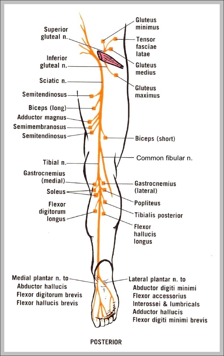
Sciatic Nerve Anatomy Diagram: A sciatic nerve anatomy diagram shows the nerve’s path from the lower spine through the pelvis and down the leg, helping diagnose and treat related pain conditions.
Nervous System Pictures

Nervous System Pictures: Pictures of the nervous system show the brain, spinal cord, and peripheral nerves, helping visualize how the body processes and transmits information.