
Tag Archives: en
Wrist Radiography Ap View Imaios Anatomy En Medical Image

The 3D images of the wrist joint and carpal bones are three-dimensional reconstructions obtained from a scanner. 340 anatomical structures of the wrist were labeled, accessible on “Anatomical parts”: General anatomy: the different regions of the wrist and hand (carpal, View Diagram Wrist Radiography Ap View Imaios Anatomy En Medical Image
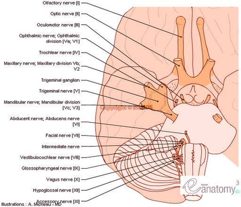
Cranial Nerves Anatomy Brainstem Human Body En Large Photo Image

Anatomy Glenohumeral Joint Shoulder Ligaments En Medical Image
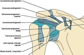
Glenohumeral ligaments (superior, middle and inferior) – the joint capsule is formed by this group of ligaments connecting the humerus to the glenoid fossa. They are the main source of stability for the shoulder, holding it in place and preventing View Diagram Anatomy Glenohumeral Joint Shoulder Ligaments En Medical Image
Muscles Arm Human Anatomy Illustration Diagram En Medical Image
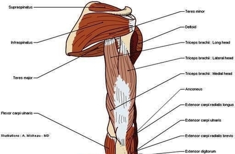
The human arm is divided into two main regions, the portion from the elbow to the wrist known as the forearm, and the segment from the shoulder to the elbow referred to as the arm. Arm muscle anatomy enables the View Diagram Muscles Arm Human Anatomy Illustration Diagram En Medical Image