Anatomy Glenohumeral Joint Shoulder Ligaments En Medical Image

This human anatomy diagram with labels depicts and explains the details and or parts of the Anatomy Glenohumeral Joint Shoulder Ligaments En Medical Image. Human anatomy diagrams and charts show internal organs, body systems, cells, conditions, sickness and symptoms information and/or tips to ensure one lives in good health.

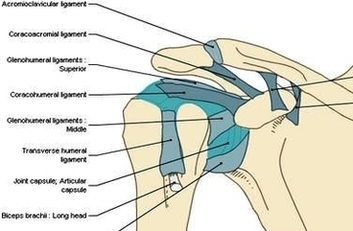
Glenohumeral ligaments (superior, middle and inferior) – the joint capsule is formed by this group of ligaments connecting the humerus to the glenoid fossa. They are the main source of stability for the shoulder, holding it in place and preventing it from dislocating anteriorly. They act to stabilise the anterior aspect of the joint.

The glenohumeral articulation involves the humeral head with the glenoid cavity of the scapula, and it represents the major articulation of the shoulder girdle.[2] The latter also includes minor articulations of the sternoclavicular (SC), acromioclavicular (AC), and scapulothoracic joints.

The shoulder ligaments are tough, elastic bands of connective tissues. They connect bone to bone, support the shoulder joints and limit their movement, playing a vital role in passive shoulder stability. The shoulder is made up of a number of different joints, and each of these joints are supported by a number of ligaments.

Anatomy Glenohumeral Joint Shoulder Ligaments En Medical Image

Anatomy Glenohumeral Joint Shoulder Ligaments En Medical Image


Tags: , , , , ,