Early Forelimb Skeleton And Flesh Image
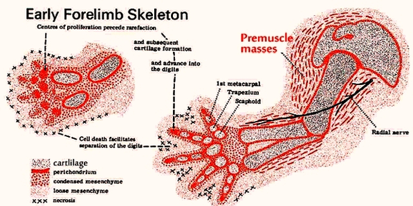
This human anatomy diagram with labels depicts and explains the details and or parts of the Early Forelimb Skeleton And Flesh Image. Human anatomy diagrams and charts show internal organs, body systems, cells, conditions, sickness and symptoms information and/or tips to ensure one lives in good health.

Anatomical landmarks: particular homologous structures on the skeleton (openings, joints, etc.) used for identifying the position of bones or other features of the anatomy. The skeleton of a dinosaur (or other vertebrate) is divided into a couple of different sections: The cranium (braincase, face, and upper jaw)

The measurement from the withers to the point of the elbow should be about the same as from the point of the elbow to the ground, ensuring adequate depth of chest. The forearm should be long and well muscled, and the cannon bone should be short with adequate flat bone. Seen from the side and front the forelimbs should be straight.

Fig. 3.3 Skeleton of the forelimb – front view The radius and ulna are equivalent to the bones of the human lower arm but, unlike the human, they are fused together to prevent the horse’s foreleg from twisting. The ulna is very small except for the olecranon process, which forms part of the elbow.

Early Forelimb Skeleton And Flesh Image

Early Forelimb Skeleton And Flesh Image


Tags: , , ,