Early Embryo Amnion Still Image

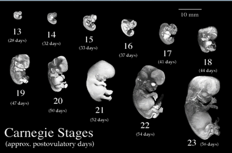
A human embryo in the sixth week after fertilization (about 41 days old and 11-14 mm long). We are looking at the developing face. The stomodeum(mouth opening) can be seen between the maxillary and mandibular processes. The nasal pits have formed nostrils.

A human embryo in the sixth week after fertilization (about 41-43 days old and 11-15 mm long). The mid-dorsal line shows the neural tube. Cranially, the embryo’s head is bent ventrally at the cervical flexure. The amnion and yolk sac are clearly seen.

Most organ development of human embryos occurs during the first 8 weeks of gestation, known as the embryonic period (see inside cover pages in Sadler). During the fetal period that succeeds it, the fetus does undergo major development but grows tremendously in size.

Early Embryo Amnion Still Image Diagram - Chart - diagrams and charts with labels. This diagram depicts Early Embryo Amnion Still Image

Early Embryo Amnion Still Image