Arm Anatomy1 Image

This human anatomy diagram with labels depicts and explains the details and or parts of the Arm Anatomy1 Image. Human anatomy diagrams and charts show internal organs, body systems, cells, conditions, sickness and symptoms information and/or tips to ensure one lives in good health.

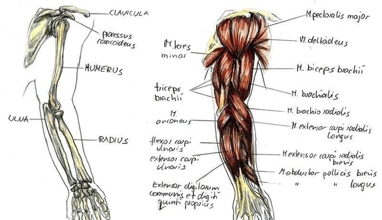
4,995 human arm anatomy stock photos and images available, or start a new search to explore more stock photos and images.

The human arm is divided into two main regions, the portion from the elbow to the wrist known as the forearm, and the segment from the shoulder to the elbow referred to as the arm. Arm muscle anatomy enables the arm to perform a variety of movements, including flexion, extension, pronation, and supination.

The muscles of the arms attach to the shoulder blade, upper arm bone ( humerus ), forearm bones ( radius and ulna ), wrist, fingers, and thumbs. These muscles control movement at the elbow, forearm, wrist, and fingers. When affected by injury or neuromuscular disorders, everyday tasks that require hand and arm use can be challenging.

Arm Anatomy1 Image

Arm Anatomy1 Image


Tags: ,