Upper Extremity Diagram1 Image

This human anatomy diagram with labels depicts and explains the details and or parts of the Upper Extremity Diagram1 Image. Human anatomy diagrams and charts show internal organs, body systems, cells, conditions, sickness and symptoms information and/or tips to ensure one lives in good health.

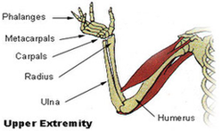
The forearm is the portion between the elbow and wrist. The thigh is the portion of the lower extremity between the hip and knee, and the calf is the portion between the knee and ankle. The normal arterial anatomy of the upper extremity is depicted graphically in Figure 13-1.

The next chapter is about the osteology of the upper limb, with all its anatomical structures, muscle insertions and ligaments of the bones: scapula, clavicle, humerus, ulna, radius, carpal bones, metacarpals and phalanges of the fingers.

The final chapter presents anatomical charts of anatomical sections of the upper limb: the axilla and the deltoid region in axial and coronal and axial sections of the arm, the elbow, forearm, wrist, carpal and metacarpal regions.

Upper Extremity Diagram1 Image

Upper Extremity Diagram1 Image


Tags: , ,