Brain Diagram1 Image

This human anatomy diagram with labels depicts and explains the details and or parts of the Brain Diagram1 Image. Human anatomy diagrams and charts show internal organs, body systems, cells, conditions, sickness and symptoms information and/or tips to ensure one lives in good health.

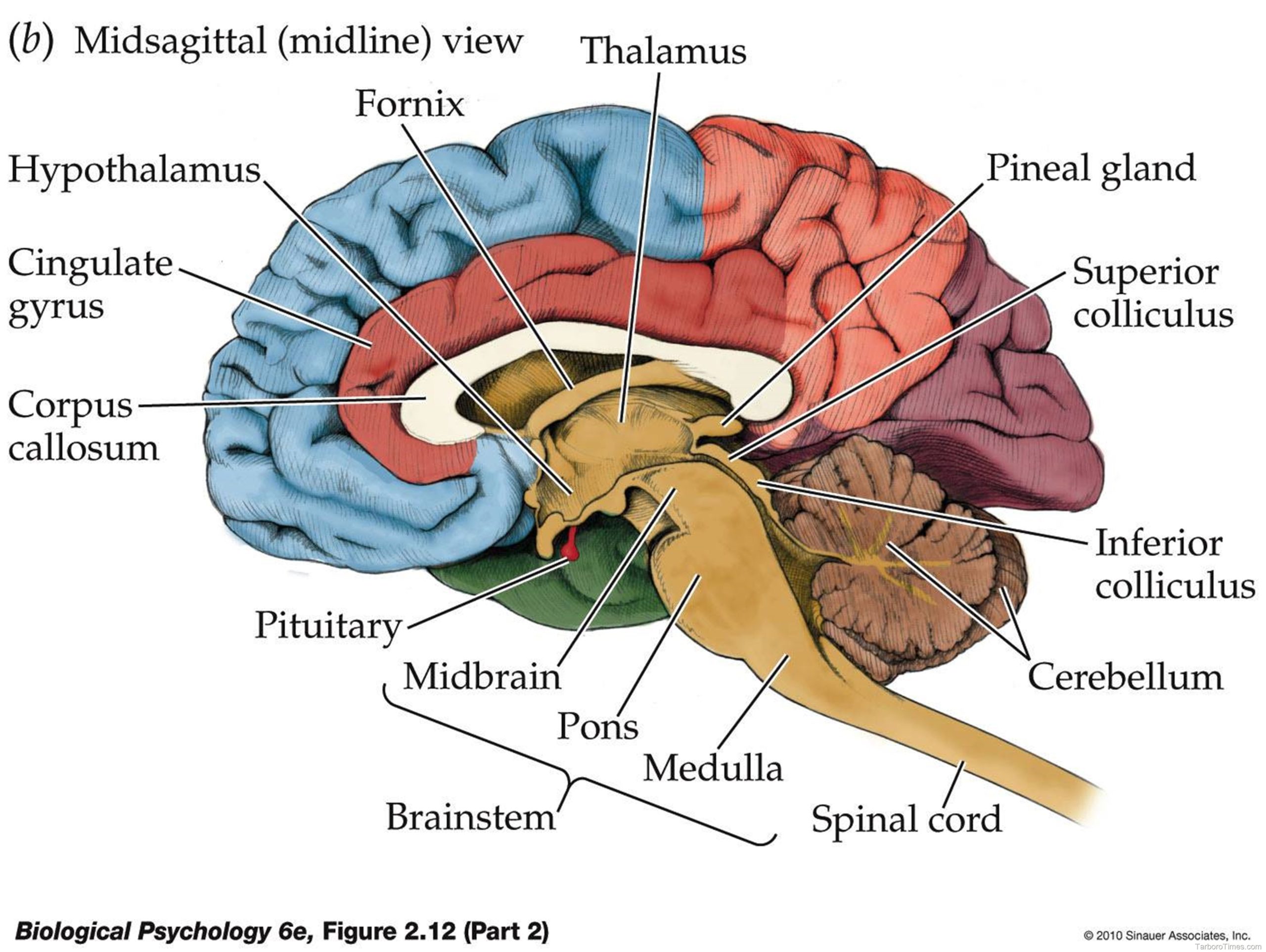
26,763 brain diagram stock photos, vectors, and illustrations are available royalty-free.

WebMD’s Brain Anatomy Page provides a detailed diagram and definition of the brain including its function, parts, and conditions that affect it. Skip to main content Check Your Symptoms Find A Doctor Find A Dentist Connect to Care Find Lowest Drug Prices Health A-Z Health A-Z Common Conditions ADD/ADHD Allergies

Labeled Diagrams of the Human Brain. Central Core. The central core consists of the thalamus, pons, cerebellum, reticular formation and medulla. These five regions are the central areas that regulate breathing, pulse, arousal, balance, sleep and early stages of processing sensory information.

Brain Diagram1 Image

Brain Diagram1 Image scaled


Tags: ,