
Neuron Diagram Image
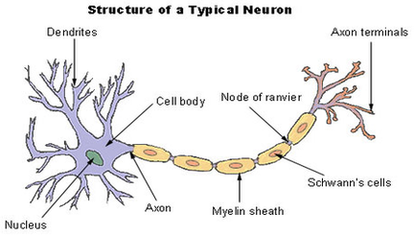
Diagram Of Neuron A neuron is a specialized cell, primarily involved in transmitting information through electrical and chemical signals. They are found in the brain, spinal cord and the peripheral nerves. A neuron is also known as the nerve cell. View Diagram Neuron Diagram Image
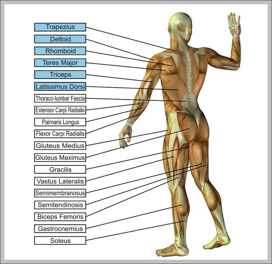
Leg Workout Routine Leg Anatomy Image

How to Design a Leg Workout Using the 15 Best Exercises. 1 1. Back squat. Target your posterior chain — or the back of your body, including the glutes and hamstrings — with a back squat. 2 2. Front squat. View Diagram Leg Workout Routine Leg Anatomy Image
Uterus Diagram Image
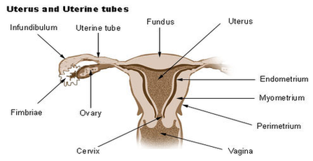
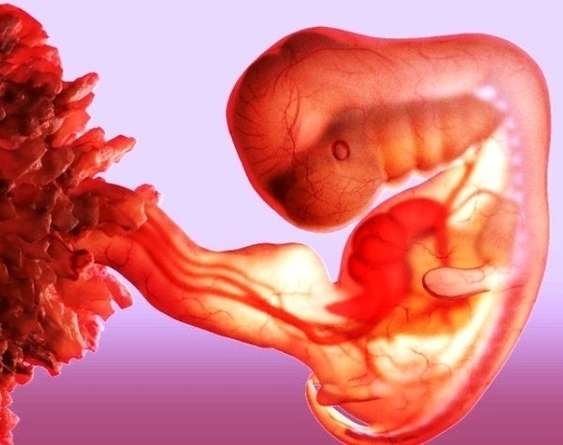
230 uterus diagram stock photos and images available or start a new search to explore more stock photos and images. An anatomical diagram depicts the method of extracting a fetus by reaching into the uterus and adjusting the baby’s position. View Diagram Uterus Diagram Image
Pull Muscles Image
A pulled muscle occurs when a muscle anywhere in the body is stretched beyond its means, leading to slight tearing of the tiny fibers that make up the muscle. A pulled muscle is sometimes known as a strained muscle, and View Diagram Pull Muscles Image
Week Fc Bbbcaelarge Image
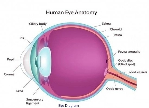
Eye Diagram Image
High Blood Pressure Image
27,768 high blood pressure stock photos, vectors, and illustrations are available royalty-free. High blood pressure, also called hypertension, is blood pressure that is higher than normal. Your blood pressure changes throughout the day based on your activities. Elevated blood pressure View Diagram High Blood Pressure Image
Foot Plantar Fasciitis Intro Image
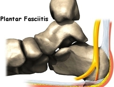
Inflammation or sprain of the tendon in the foot, heel spur, bursitis. plantar fasciitis stock pictures, royalty-free photos & images Woman suffering from heel pain. Inflammation or sprain of the… The concept of diseases and pains in the leg. Woman View Diagram Foot Plantar Fasciitis Intro Image
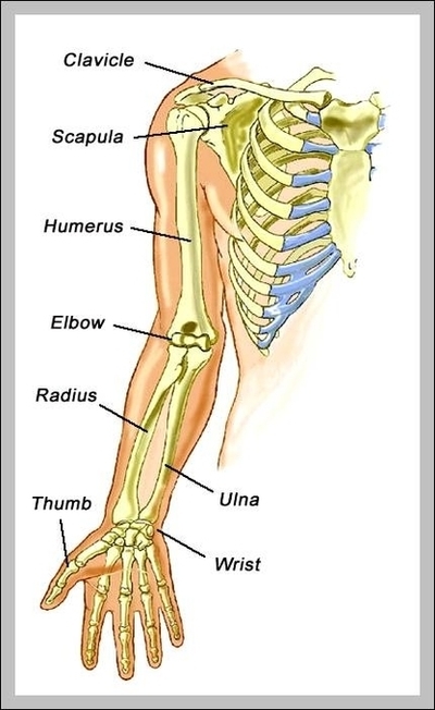
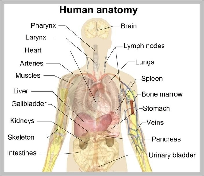
Human Anatomy Chart Image
5,915 human anatomy diagram stock photos and images available or search for the human anatomy or anatomy model to find more great stock photos and pictures. Anatomy charts can be specific to one part of the body, such as a View Diagram Human Anatomy Chart Image
Turkey Avocado Blt Paneratures Image

Panera Turkey Avocado BLT (Copycat) Cut bread in half and put a generous amount of mayo on each piece. For each sandwich use 5 slices of turkey, 1 1/2 pieces of bacon, 1/2 of an avocado (about 4 slices), 2 View Diagram Turkey Avocado Blt Paneratures Image
Anatomy Of Human Body Picture Image
29,336 human body anatomy stock photos and images available, or search for human body anatomy vector or the human body anatomy body to find more great stock photos and pictures. Abstract image human body in the form of a starry View Diagram Anatomy Of Human Body Picture Image
Diagram Of Nasa Middle Ear Image
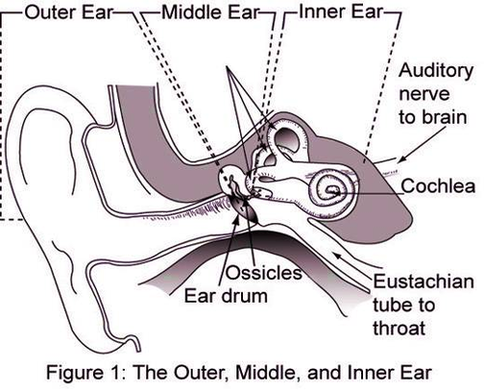
Also known as the tympanic cavity, the middle ear is an air-filled, membrane-lined space located between the ear canal and the Eustachian tube, cochlea, and auditory nerve. The eardrum separates this space from the ear canal. The Structure of Human View Diagram Diagram Of Nasa Middle Ear Image
Anatomy And Physiology Of Animals Growing Bone1 Image
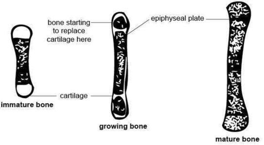
138Anatomy and physiology of domestic animals associated with bone formation, it can occur in other tissues. There are two general classes of bone formation. Intramembranous ossifi cation occurs when bone develops from a fi brous membrane. The fl at bones View Diagram Anatomy And Physiology Of Animals Growing Bone1 Image
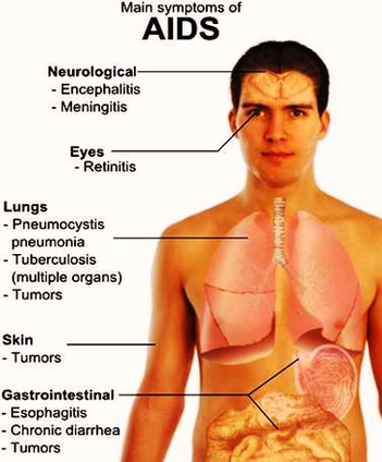
Hiv Aids Vaccine Image

Nutrition And Food Labeltures Image
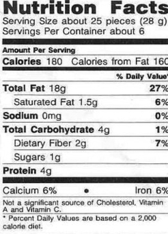
Labels show calories, fat content, cholesterol, carbohydrates, protein, sodium, sugars, daily recommended litmits, vitamins and specfic ingredients in the foods. Prominent top label shows a whopping 680 mg soldium in the food product. nutrition label stock pictures, royalty-free photos & View Diagram Nutrition And Food Labeltures Image
Female Body Image
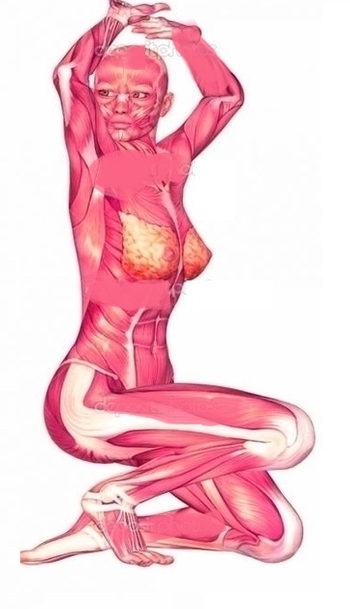
Diagram Asthma Attack Anatomy Image
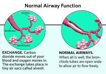
Asthma_0312 Slideshow During an asthma attack, muscles around the airways tighten, and the airway linings swell. Excess mucus secretion is produced in the airways that can block the air tubes and lungs. When air is trapped, breathing becomes difficult. Excess View Diagram Diagram Asthma Attack Anatomy Image
Anatomy Of Human Body Organs Image
12,239 human body anatomy organs stock photos and images available or start a new search to explore more stock photos and images. Human internal organs vector Vector isolated illustration of human internal organs in male body. Stomach, liver, intestine, bladder, View Diagram Anatomy Of Human Body Organs Image
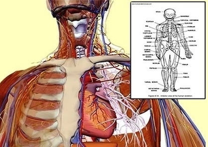
Anatomy Of The Throat Image
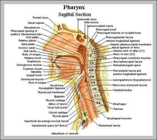
Understanding the Basics of Throat anatomy The throat is one of the most complex parts of the human body. It starts from the pharynx and extends to the upper end of the esophagus. Immediately following the pharynx are the larynx, View Diagram Anatomy Of The Throat Image