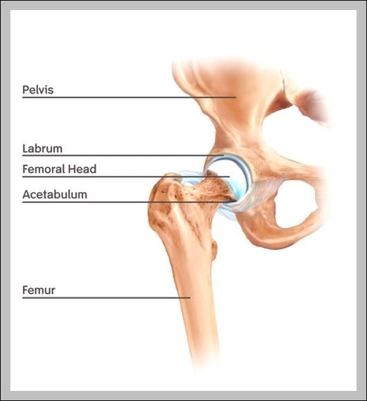
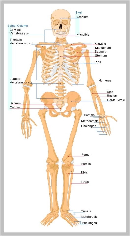
The Longest Muscle In The Body

The sartorius muscle (/sɑːrˈtɔːriəs/) is the longest muscle in the human body. It is a long, thin, superficial muscle that runs down the length of the thigh in the anterior compartment. The sartorius muscle (/sɑːrˈtɔːriəs/) is the longest muscle in View Diagram The Longest Muscle In The Body