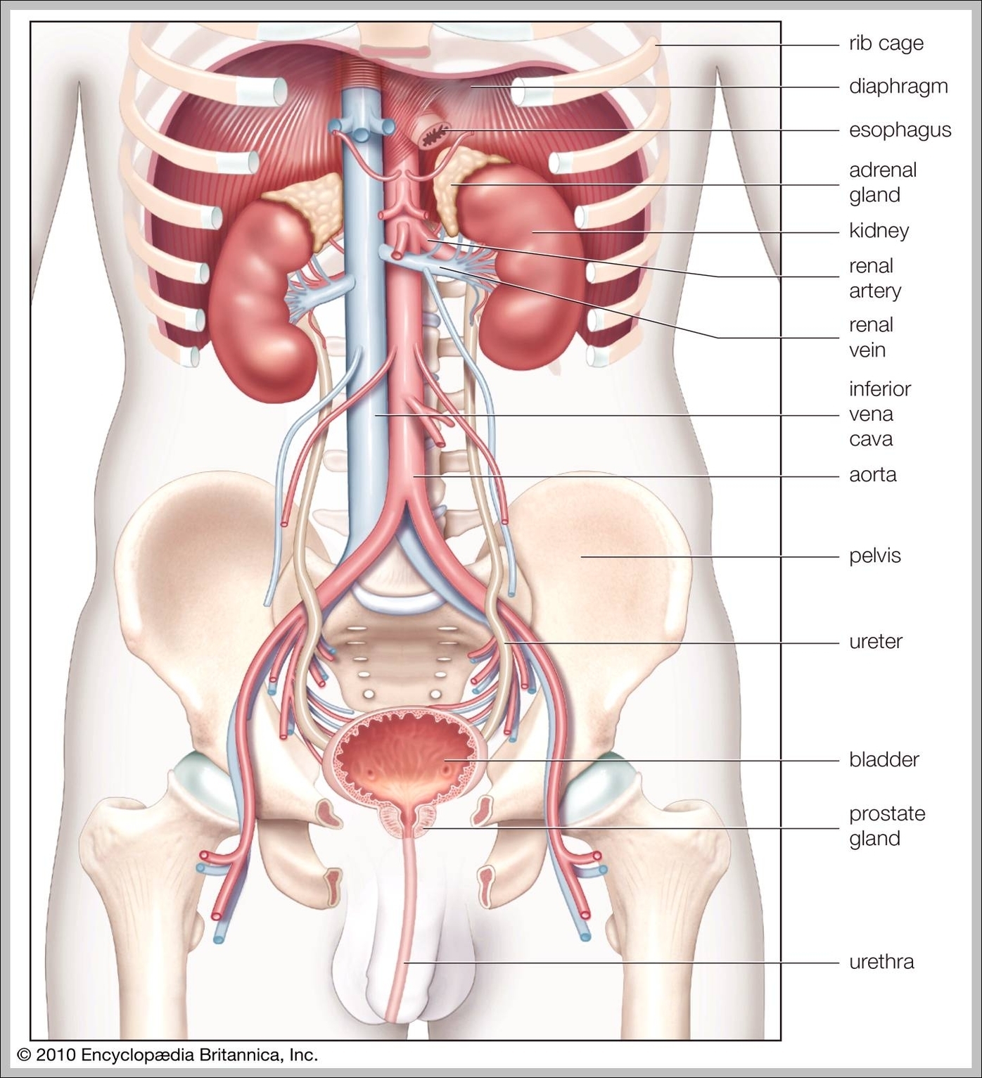
Pictures Of Skeletal System
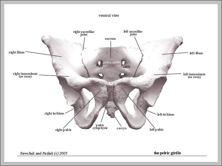
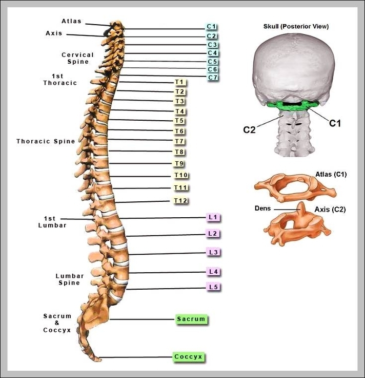
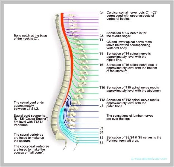
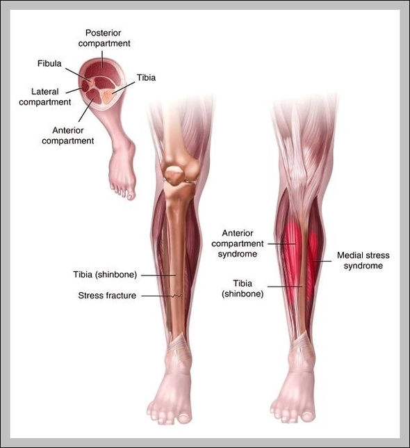
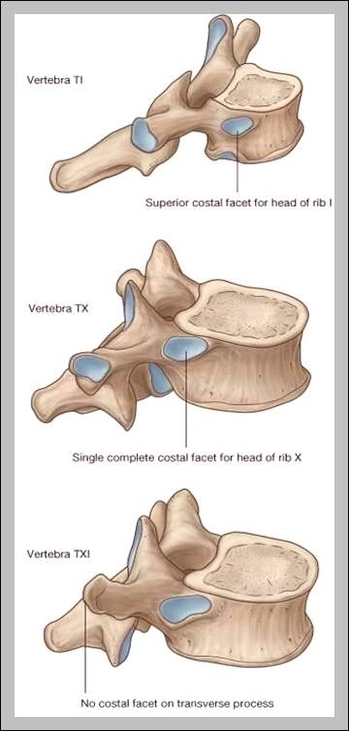
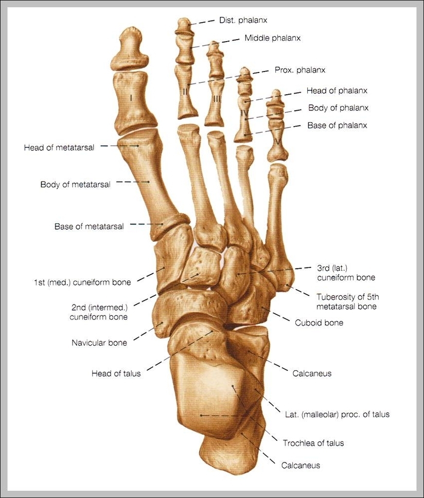
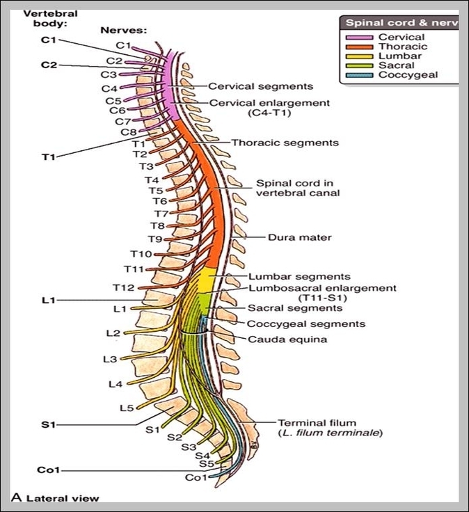
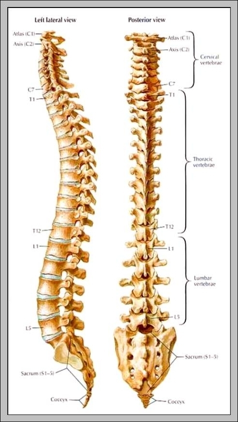
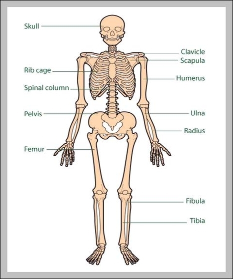
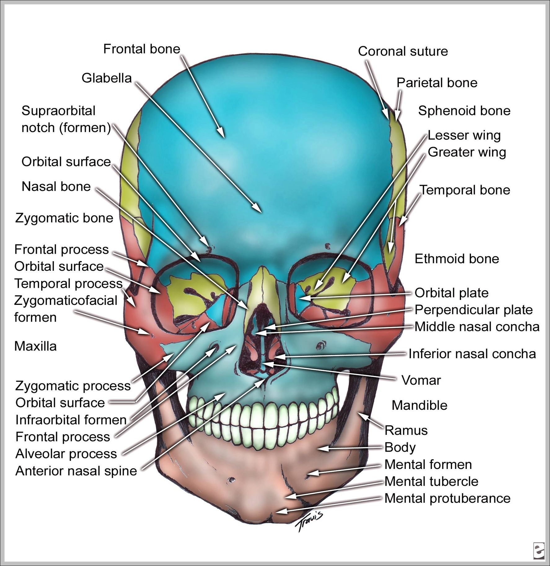
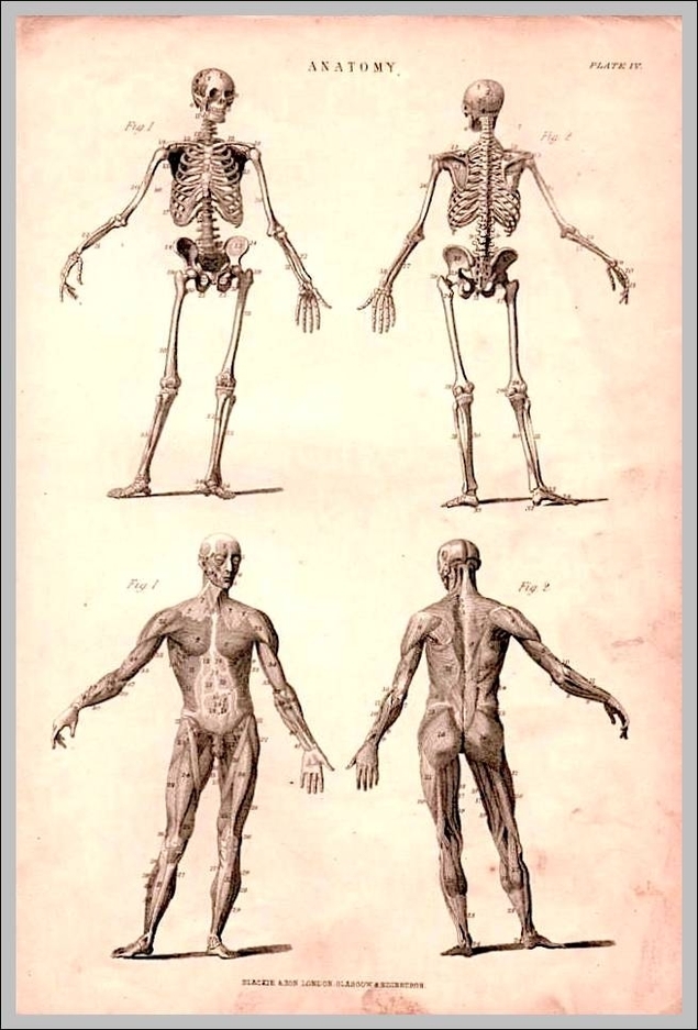
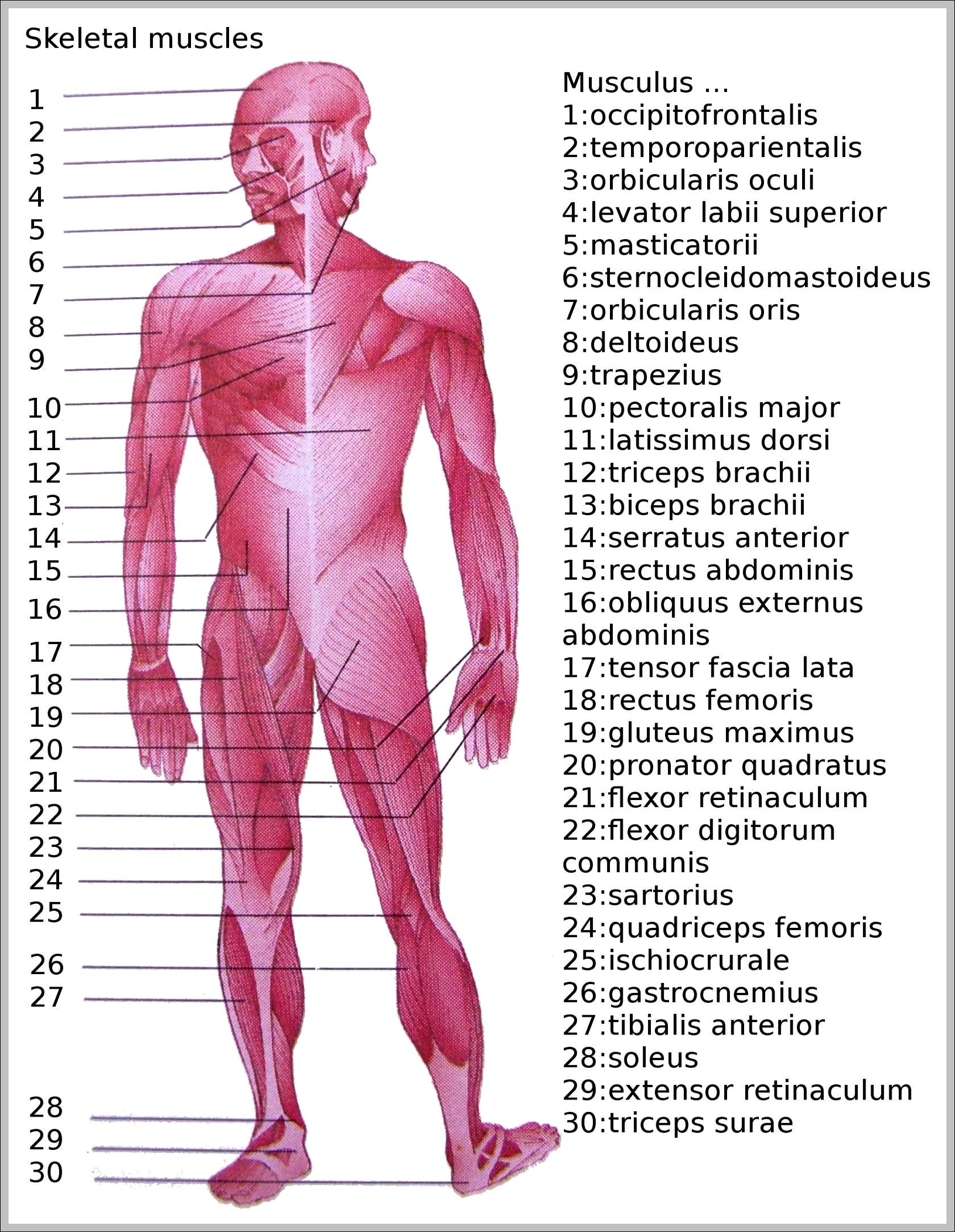
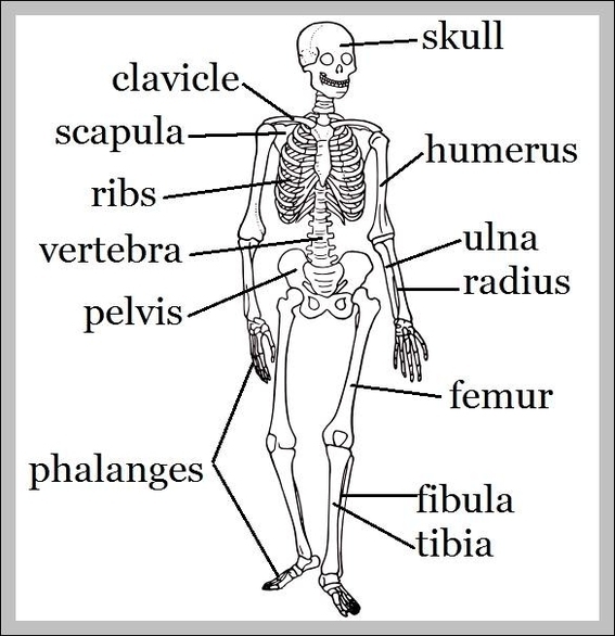
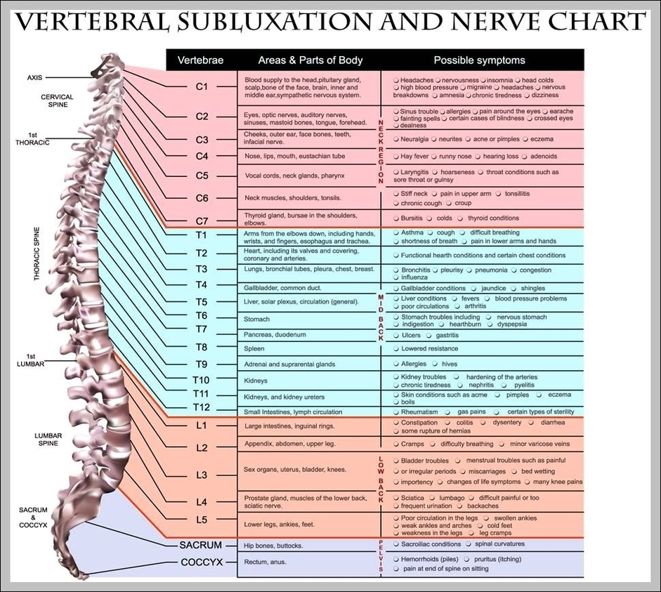
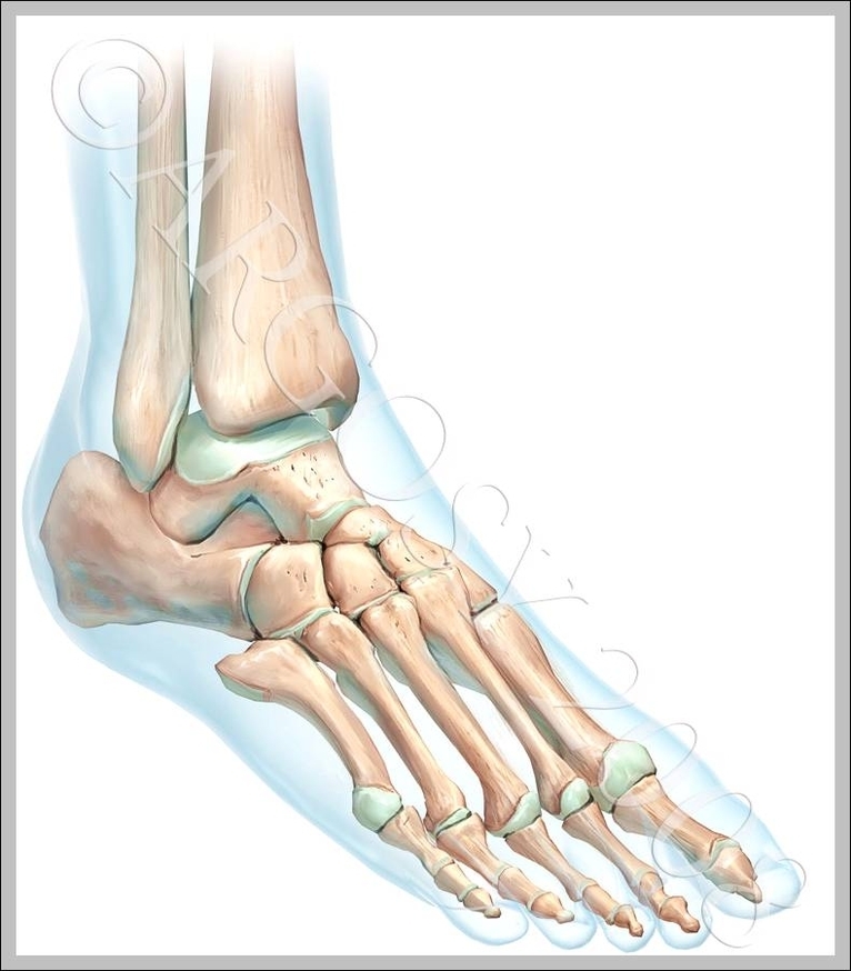
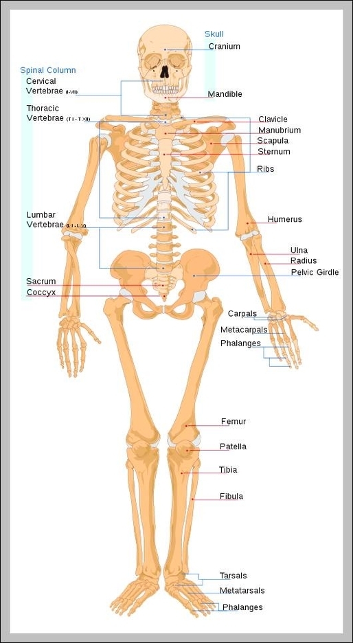
Skeletal System 1 Skull. 2 Hyoid and Auditory Ossicles. 3 Vertebrae. 4 Ribs and Sternum. 5 Pectoral Girdle and Upper Limb. 6 Pelvic Girdle and Lower Limb. 7 Microscopic Structure of Bones. 8 Types of Bones. 9 Parts of Bones. View Diagram Pictures Of Skeletal System