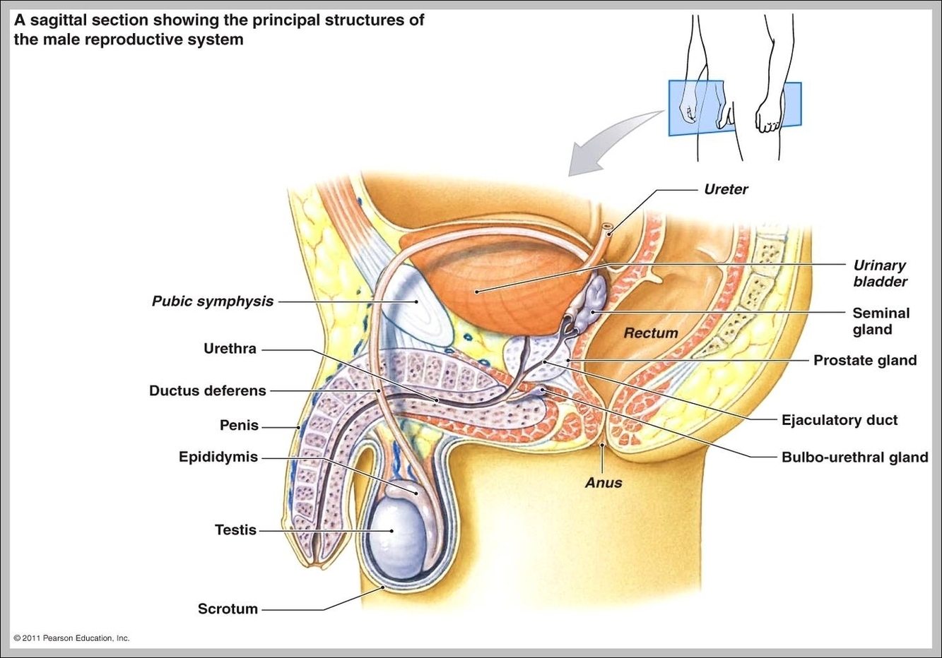

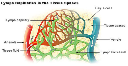

Ultrasoud Weeks Image

All about normal 10 week ultrasound. All about your normal 9 week ultrasound. Sharing is caring! Normal 13 week baby ultrasound. First let me tell you that by now you probably have all your first trimester regular ultrasounds done. The View Diagram Ultrasoud Weeks Image