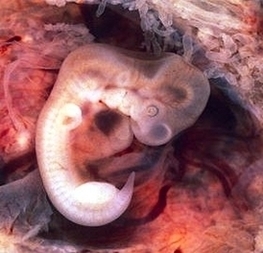
Px Tubal Pregnancy With Embryo Image

This human anatomy diagram with labels depicts and explains the details and or parts of the Px Tubal Pregnancy With Embryo Image. Human anatomy diagrams and charts show internal organs, body systems, cells, conditions, sickness and symptoms information and/or tips to ensure one lives in good health.

Tubal pregnancy: A pregnancy that is not in the usual place within the uterus but is located in the Fallopian tube. Tubal pregnancies are due to the inability of the fertilized egg to make its way through the Fallopian tube into the uterus.

Tubal pregnancies are most often diagnosed in the sixth through the ninth weeks of pregnancy. 5 Your healthcare provider will be able to diagnose a tubal pregnancy after a medical exam and other testing, which may include: 6 Pelvic examination to test for pain, tenderness, or a mass in the abdomen

Pregnancy tests taken at the beginning of a tubal pregnancy will usually show a positive result, even though the pregnancy won’t be able to grow into a healthy baby. Some people may experience no symptoms at first, but as the fertilized egg continues to grow in the fallopian tube, there can be some serious and more noticeable signs, including: 2

Px Tubal Pregnancy With Embryo Image

Px Tubal Pregnancy With Embryo Image


Tags: , , , ,