Picture Of Neck Muscles

picture of neck muscles

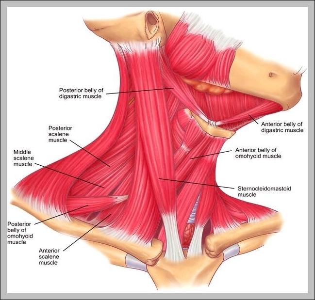
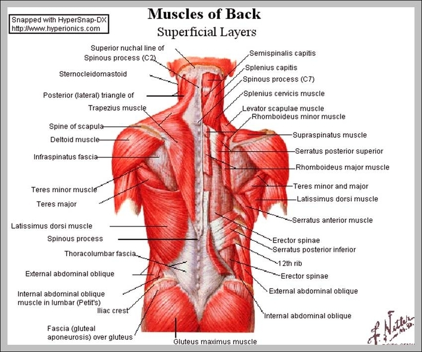
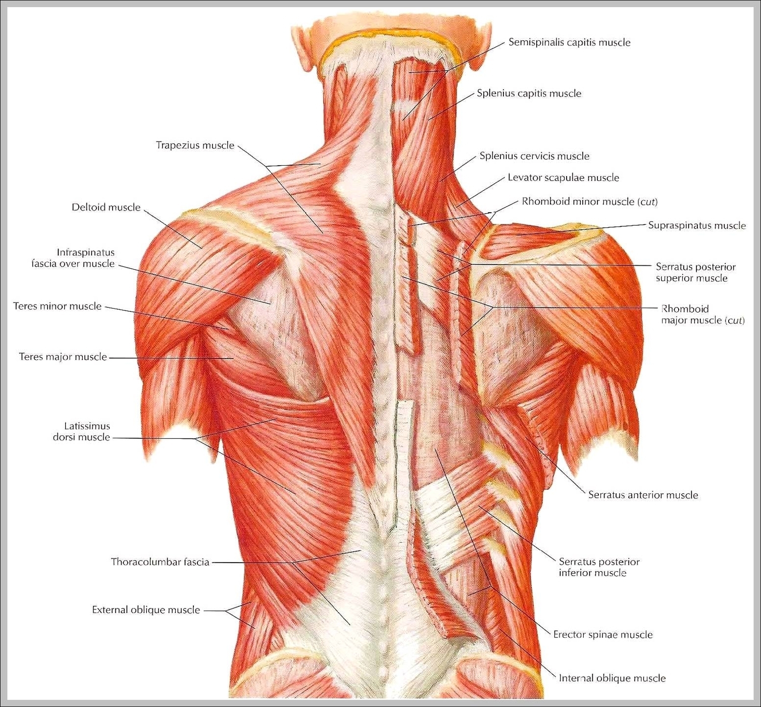
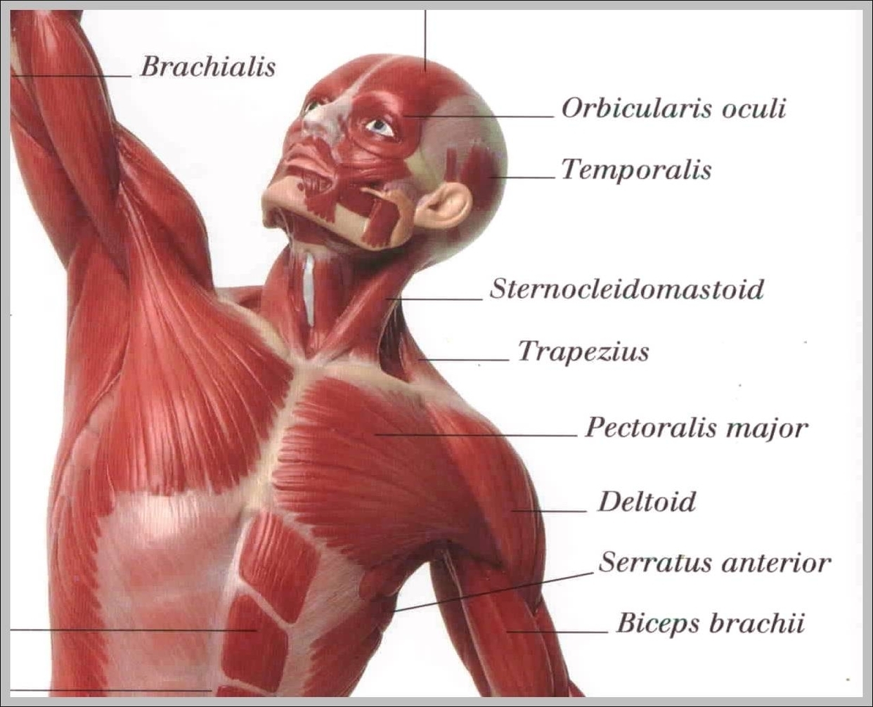
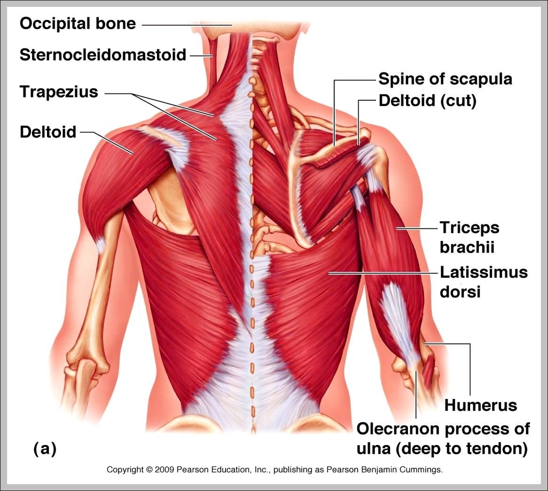
Muscles of neck. The muscles of the neck run from the base of the skull to the upper back and work together to bend the head and assist in breathing. The motion of the muscles of the neck are divided View Diagram Picture Of Neck Muscles

What Are Muscle Cells

what are muscle cells

Types of muscle cells Skeletal Muscle cells. Skeletal muscle cells are long, cylindrical, and striated. Cardiac Muscle Cells. Cardiomyocytes are short and narrow, and fairly rectangular in shape. Smooth Muscle Cells. Smooth muscle cells are spindle-shaped and contain a single View Diagram What Are Muscle Cells

Picture Of Human Leg Muscles

picture of human leg muscles

You may also find Vastus intermedius deep layer, Vastus medial, Adductor brevis m., Adductor longus m., Adductor Magnus & minimus m., Sartorius m., Gracilis m., Semimembranous muscle, Soleus m., Gastrocnemius m., Plantaris m. in it. We are pleased to provide View Diagram Picture Of Human Leg Muscles

Upper Back Muscle Diagram

upper back muscle diagram

Common causes of upper back pain 1. Muscle deconditioning and poor posture. Using the muscles infrequently or incorrectly can cause back pain. People can… 2. Muscle overuse. Overusing back muscles is another common cause of upper back pain. This typically View Diagram Upper Back Muscle Diagram

Picture Of Muscles In Back

picture of muscles in back

The back anatomy includes the latissimus dorsi, trapezius, erector spinae, rhomboid, and the teres major. On this page, you’ll learn about each of these muscles, their locations and functional anatomy. Function of the Back Muscles There are several individual muscles View Diagram Picture Of Muscles In Back

Picture Of Human Skeleton With Muscles

picture of human skeleton with muscles

There are around 640 skeletal muscles within the typical human body. There are around 640 skeletal muscles within the typical human body. Almost every muscle constitutes one part of a pair of identical bilateral muscles, found on both sides, resulting View Diagram Picture Of Human Skeleton With Muscles

Types Of Muscles

types of muscles

There are three types of muscle found in the human body: Skeletal Muscle. Cardiac Muscle (heart muscle) Skeletal muscle. Skeletal Muscles are those which attach to bones and have the main function of contracting to facilitate movement of our skeletons. View Diagram Types Of Muscles

Picture Of Deltoid Muscle

picture of deltoid muscle

The deltoid muscles are shoulder muscles of the human body. These muscles are broken into three groups, which are the anterior, lateral, and posterior muscles. The anterior deltoid is the muscle located on the front area of the shoulder. This View Diagram Picture Of Deltoid Muscle

Skeletal Muscle Picture

skeletal muscle picture

Skeletal muscles are the muscles that we can see and feel through our skin. An individual skeletal muscle is considered an organ within the muscular system of the body. The skeletal muscle works with nerve tissue, connective tissue and vascular View Diagram Skeletal Muscle Picture

Shoulder Muscle Names

shoulder muscle names

The shoulder girdle muscles make the scapula (shoulder blade) move. Muscles of the shoulder joint include the subscapularis, latissimus dorsi, infraspinatus, teres minor, teres major, supraspinatus, deltoid, and pectoralis major (pec muscles). On this page: For that reason, and because View Diagram Shoulder Muscle Names

Picture Of Back Muscles

picture of back muscles

Back Anatomy: All About the Back Muscles 1 Function of the Back Muscles 2 Latissimus Dorsi (Lats) 3 Trapezius (Traps) 4 Erector Spinae (Spinal Erectors) 5 Rhomboid 6 Teres Major Lower Back and Superficial Muscles The muscles of the lower View Diagram Picture Of Back Muscles

Visceral Muscles

visceral muscles

…the vertebrate musculature are the visceral musculature and the somatic musculature (the striated muscles of the body wall). Somatic musculature may be divided into appendicular, or limb, muscles and axial muscles. The axial muscles include the muscles of the tail, View Diagram Visceral Muscles

Torso Muscle Diagram

torso muscle diagram

Muscle Spasms in Torso: Causes and Treatments 1 Spasms felt throughout the chest and abdomen region can be caused by… 2 Pulled Muscle in the Chest. A pulled muscle in the chest can result in muscle spasms. 3 Injury to View Diagram Torso Muscle Diagram

Visceral Smooth Muscle

visceral smooth muscle

Single-unit smooth muscle, or visceral smooth muscle is a type of smooth muscle found in the uterus, gastro-intestinal tract, and the bladder. In SUVSM, a single smooth muscle cell in a bundle is innervated by an autonomic nerve fiber. Visceral View Diagram Visceral Smooth Muscle

Upper Body Muscle

upper body muscle

List of Upper Body Muscles Neck and Shoulders. The neck primarily consists of the sternocleidomastoid and the splenius muscles,… Chest. The chest region includes two muscle groups and one individual muscle. Abdominals. The abdominals primarily consist of the rectus abdominis, View Diagram Upper Body Muscle

The Longest Muscle In The Body

the longest muscle in the body

The sartorius muscle (/sɑːrˈtɔːriəs/) is the longest muscle in the human body. It is a long, thin, superficial muscle that runs down the length of the thigh in the anterior compartment. The sartorius muscle (/sɑːrˈtɔːriəs/) is the longest muscle in View Diagram The Longest Muscle In The Body

Upper Body Muscle Anatomy

upper body muscle anatomy

The major muscles in the upper torso of the body include: Trapezius: This muscle extends across the neck, shoulder, and back. Rhomboid major: Attached to the shoulder blade, this muscle is one of many that aids shoulder movement. Infraspinatus: This View Diagram Upper Body Muscle Anatomy

Skeletal Muscle Labeled

skeletal muscle labeled

Skeletal Muscle Labeled: A skeletal muscle labeled diagram highlights the major skeletal muscles in the body, such as the quadriceps, biceps, and deltoids, along with their names and locations.