Dorsal Forearm Diag Flat Image

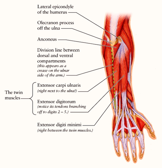
Anatomy of the distal forearm 1 Soft tissue anatomy – dorsal. 2 Soft tissue anatomy – palmar. 3 Bony anatomy. 4 Principle of columns. The distal forearm may be thought of in terms of three columns. The ulna forms one column. The… More …

Currently, computed tomography (CT) and magnetic resonance imaging (MRI) play an important role in evaluation of injuries to the distal forearm, wrist, and hand (see the text that follows).

Volar/dorsal splints are splints that extend from the middle of the forearm to the distal palmar crease (top crease in the palm). Volar splints are applied to the palm side of the forearm, wrist and hand, while dorsal splints are applied to the top side of the forearm, wrist, and hand.

Dorsal Forearm Diag Flat Image Diagram - Chart - diagrams and charts with labels. This diagram depicts Dorsal Forearm Diag Flat Image

Dorsal Forearm Diag Flat Image