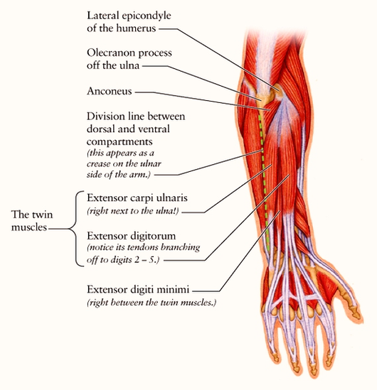
Diagram Dorsal Forearm Diagflat Image
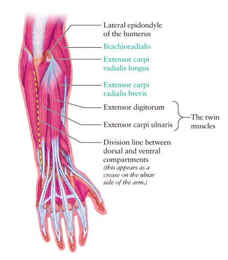
Muscles Just like the arm, the forearm is divided into two compartments by deep fascia; the interosseous membrane, and the fibrous intermuscular septa. This creates an anterior compartment that contains the flexor muscles, and a posterior one that contains the View Diagram Diagram Dorsal Forearm Diagflat Image