Skip to content

Human Anatomy Diagrams

Menu

Primary menu

  • Top
  • Anatomy
  • Muscles
  • Systems
  • Body Parts
  • Organs
  • Cells
  • Bones
  • Teeth
  • Cancer
  • Health

Labeled Diagram Of Human Heart Image

Posted on June 8, 2022 by anatomy
Labeled Diagram Of Human Heart Image

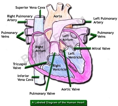
Human Heart Diagram Labeled. The human heart is an organ responsible for pumping blood through the body, moving the blood (which carries valuable oxygen) to all the tissues in the body. Without the heart, the tissues couldn’t get the oxygen View Diagram Labeled Diagram Of Human Heart Image→

Posted in Anatomy | Tagged heart, labeled

Cancer Stem Cells Diagram Image

Posted on June 8, 2022 by anatomy
Cancer Stem Cells Diagram Image

The cancer stem cell model. The cancer stem cell model, also known as the Hierarchical Model proposes that tumors are hierarchically organized (CSCs lying at the apex (Fig. CSCs may generate tumors through the stem cell processes of self-renewal and View Diagram Cancer Stem Cells Diagram Image→

Posted in Cancer, Cells | Tagged cancer, cells, stem

Lymph Capillary Diagram Image

Posted on June 8, 2022 by anatomy
Lymph Capillary Diagram Image

Lymph capillaries or lymphatic capillaries are tiny, thin-walled microvessels located in the spaces between cells (except in the central nervous system and non-vascular tissues) which serve to drain and process extracellular fluid. Upon entering the lumen of a lymphatic capillary, View Diagram Lymph Capillary Diagram Image→

Posted in Cells | Tagged capillary, lymph

Dental Crown Tooth Before Image

Posted on June 8, 2022 by anatomy
Dental Crown Tooth Before Image

Dental crowns plays an important role in supporting and restoring the normal structure and appearance of damaged teeth. If any of your front teeth has suffered severe damage, broken or extensively decayed, a dental crown repair is usually recommended. However, View Diagram Dental Crown Tooth Before Image→

Posted in Anatomy | Tagged Before, crown, dental, tooth

Gestational Diabetes Symptoms Ukwhfqbi Image

Posted on June 8, 2022 by anatomy
Gestational Diabetes Symptoms Ukwhfqbi Image

Some pregnant women do notice subtle signs of gestational diabetes. The symptoms are similar to those of other forms of diabetes. But they’re also common symptoms in all pregnant women, so they’re easy to miss as the sign that something’s View Diagram Gestational Diabetes Symptoms Ukwhfqbi Image→

Posted in Anatomy | Tagged diabetes, gestational, symptoms, ukwhfqbi

Hiv Aids Chrt Photo Image

Posted on June 8, 2022 by anatomy

Hiv Aids Chrt Photo Image

Posted in Anatomy | Tagged aids, chrt, hiv, photo

Adult Foot Fx Anatomya Image

Posted on June 8, 2022 by anatomy

Adult Foot Fx Anatomya Image

Posted in Anatomy | Tagged a, adult, foot, fx

Human Ear Diagram Image

Posted on June 8, 2022 by anatomy
Human Ear Diagram Image

The Structure of Human Ear. Helix: It is the prominent outer rim of the external ear. Antihelix: It is the cartilage curve that is situated parallel to the helix. Crus of the Helix: It is the landmark of the outer View Diagram Human Ear Diagram Image→

Posted in Anatomy | Tagged ear, human

Motor Sensory Image

Posted on June 8, 2022 by anatomy
Motor Sensory Image

Motor imagery is the mental execution of a movement without any overt movement or without any peripheral (muscle) activation. It has been shown that motor imagery leads to the activation of the same brain areas as actual movement. Information from View Diagram Motor Sensory Image→

Posted in Anatomy | Tagged motor, sensory

Costovertebral Joints Anatomy Diagram Image

Posted on June 8, 2022 by anatomy
Costovertebral Joints Anatomy Diagram Image

The costovertebral joints describe two groups of synovial plane joints which connect the proximal end of the ribs with their corresponding vertebrae, enclosing the thoracic cage from the posterior side. Precisely, these joints are described as; The costovertebral complex is View Diagram Costovertebral Joints Anatomy Diagram Image→

Posted in Anatomy | Tagged costovertebral, joints

Footankle Bony Anat Image

Posted on June 8, 2022 by anatomy
Footankle Bony Anat Image

This article outlines the basic anatomy of the foot bones, along with some of the most common conditions affecting these bones. The human foot consists of 26 bones. These bones fall into three groups: the tarsal bones, metatarsal bones, and View Diagram Footankle Bony Anat Image→

Posted in Anatomy | Tagged anat, bony, footankle

Diagram Of Druker What Causes Blood Cancer Image

Posted on June 8, 2022 by anatomy
Diagram Of Druker What Causes Blood Cancer Image

Blood cancers occur when abnormal blood cells start growing out of control, interrupting the function of normal blood cells, which fight off infection and produce new blood cells. The three main types of blood and bone marrow cancer are leukemia, View Diagram Diagram Of Druker What Causes Blood Cancer Image→

Posted in Cancer, Cells | Tagged blood, Causes, druker, What

Red White Blood Cells Image

Posted on June 8, 2022 by anatomy

Red White Blood Cells Image

Posted in Cells | Tagged blood, cells, red, white

Aterial Pressure Fluctuations Image

Posted on June 8, 2022 by anatomy
Aterial Pressure Fluctuations Image

Arterial pressure results from the pressure exerted by the blood in the large arteries. Blood pressure depends on cardiac output and total peripheral resistance. Arterial pressure fluctuates with each heart beat, according to the pumping of the heart. Moreover, a View Diagram Aterial Pressure Fluctuations Image→

Posted in Anatomy | Tagged aterial, fluctuations, pressure

Diabetes Glucose Testings Image

Posted on June 8, 2022 by anatomy
Diabetes Glucose Testings Image

The A1C test measures your average blood sugar level over the past 2 or 3 months. An A1C below 5.7% is normal, between 5.7 and 6.4% indicates you have prediabetes, and 6.5% or higher indicates you have diabetes. Fasting Blood View Diagram Diabetes Glucose Testings Image→

Posted in Anatomy | Tagged diabetes, glucose, testings

Human Body Diagrams Image

Posted on June 8, 2022 by anatomy

Human Body Diagrams Image

Posted in Anatomy | Tagged body, human, s

Male Reproductive System Anatomy Image

Posted on June 8, 2022 by anatomy

Male Reproductive System Anatomy Image

Posted in System | Tagged male, reproductive, system

Power Torso Overview Image

Posted on June 8, 2022 by anatomy

Power Torso Overview Image

Posted in Anatomy | Tagged overview, power, torso

Map Of The Human Body Organs Image

Posted on June 8, 2022 by anatomy
Map Of The Human Body Organs Image

12,251 human body organs anatomy stock photos and images available, or start a new search to explore more stock photos and images. Human internal organs vector Vector isolated illustration of human internal organs in male body. Stomach, liver, intestine, bladder, View Diagram Map Of The Human Body Organs Image→

Posted in Anatomy, Organs | Tagged body, map

Gluten Free Diagram Image

Posted on June 8, 2022 by anatomy

Gluten Free Diagram Image

Posted in Anatomy | Tagged free, gluten

Post navigation

← Older posts
Newer posts →

Primary Sidebar Widget Area

Top Diagrams

  • Human Body Organ Locations Human Body Organ Locations

  • Map Of Body Muscles Map Of Body Muscles

  • Human Organs Back View Human Organs Back View

  • Female Abdominal Anatomy Female Abdominal Anatomy

  • Male Anatomy Pictures Male Anatomy Pictures

  • Female Muscular System Female Muscular System

  • Map Of Human Organs Map Of Human Organs

  • Female Human Anatomy Organs Female Human Anatomy Organs

  • Male Body Organs Diagram Male Body Organs Diagram

  • Picture Of Organs On Left Side Of Body Picture Of Organs On Left Side Of Body

  • Lower Back Hip Anatomy Lower Back Hip Anatomy

  • Muscular System Full Body Diagram Muscular System Full Body Diagram

  • 12 Organ Systems 12 Organ Systems

  • Lower Back Anatomy Pictures Lower Back Anatomy Pictures

  • Pictures Of The Heart With Labels Pictures Of The Heart With Labels

Latest Posts

  • Varicose veins
  • The Stages of Walking
  • Quadratus Lumborum
  • Lateral View of the Scapula Glenoid Fossa Acromion and Coronoid Process Labelled
  • Anatomy of the Origin of the Trigeminal Nerve Nuclei and Ganglia
  • Structure of the Hair Follicle
  • Brain stem
  • Lymphatic Trunks and Ducts System
  • Diagram of Spinal Cord Outflow Creating Posterior and Anterior Rami
  • Relationship between the Ureter and the Uterine Artery
  • Anatomical Course and Branches of the Pudendal Nerve
  • Types of Spina Bifida
  • Posterior Cutaneous Nerve of Thigh
  • X Ray Generation Imagine
  • Muscles Innervated by the Accessory Nerve

Categories

  • Anatomy
  • Body Parts
  • Bones
  • Cancer
  • Cells
  • Health
  • Muscles
  • Organs
  • System
  • Teeth
  • Top

Top Tags

  • muscles (205)
  • muscle (171)
  • human (156)
  • human muscles (138)
  • muscles diagram (138)
  • muscles anatomy (138)
  • human muscle diagram (136)
  • muscular system (136)
  • body (119)
  • system (119)
  • body organs (119)
  • blood (82)
  • parts of a human body (77)
  • human body parts (77)
  • body parts (77)
  • organs (67)
  • organ (67)
  • pregnancy (66)
  • diabetes (62)
  • organ anatomy (62)
Copyright © 2026 . All Rights Reserved.
Theme: Catch Box by Catch Themes
Scroll Up