
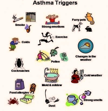
Gastroparesis Dia Stomach Image
Gastroparesis. Gastroparesis is a disease in which the stomach cannot empty itself of food in a normal fashion. Symptoms include heartburn, nausea, vomiting, and feeling full quickly when eating. Treatments include medications and possibly surgery. Gastroparesis is also often referred View Diagram Gastroparesis Dia Stomach Image
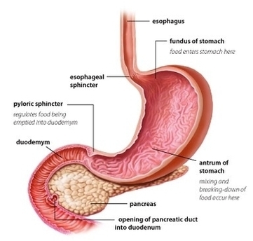
Clean Air House Diagram Image
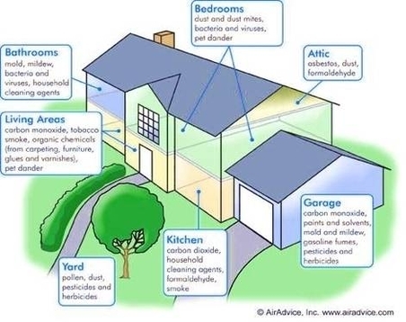
Diagram Diab Symp Image
Our circuit diagram symbol library is schematic and includes many icons commonly used by engineers. From transistors to logic gates, you’ll find icons that are modeled to international standards. At US, the diaphragm appears as a thick echogenic line. M-mode View Diagram Diagram Diab Symp Image
Body Organs Pictures Image
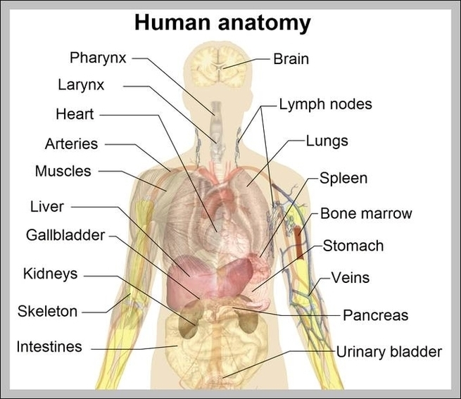
Diagram Of Week Pregnancy Image
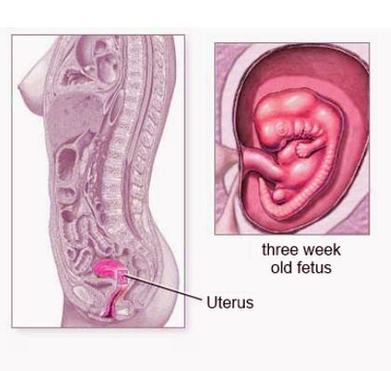
This week by week pregnancy chart covers fetal development from 4 weeks to 42 weeks – and it lets you know what pregnancy signs and symptoms you might experience that week. The chart also shows a picture of what your View Diagram Diagram Of Week Pregnancy Image
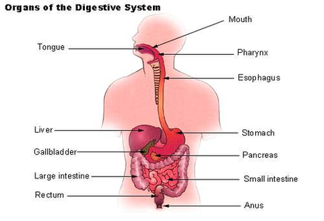
Digestive System Image
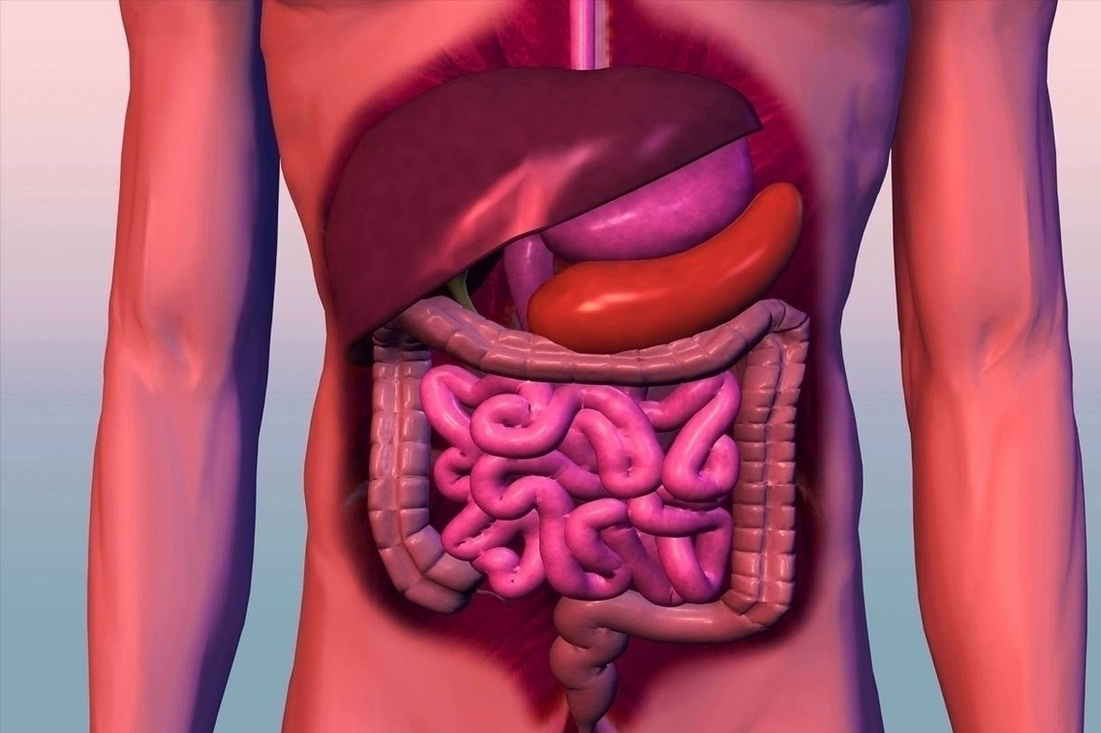
125,099 human digestive system stock photos and images available, or search for human digestive system illustration or human digestive system anatomy to find more great stock photos and pictures. About Us Search Term Digestive System By: Tim Taylor Last Updated: View Diagram Digestive System Image
Aids Virus Image

Diagram Crukmig Image
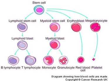
Diagram Of Heart Blood Flow Image
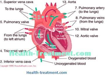
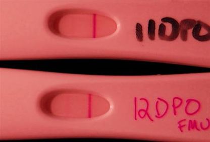
Diagram Faint Positive Pregnancy Tests Di Mitra Sama Keluarga Image
Anatomy Human Brain Image
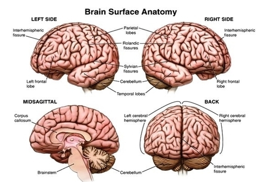
10,526 brain anatomy stock photos and images available, or search for human brain anatomy or brain anatomy illustration to find more great stock photos and pictures. The anatomy of the human brain it is characterized by the following parts: Cerebral View Diagram Anatomy Human Brain Image
Embryo Days Weeks Image

Your embryo has completed the most critical portion of development. Their skin is still translucent, but their tiny limbs can bend and fine details like nails are starting to form. Read about your pregnancy at 10 weeks. What You’re Seeing: View Diagram Embryo Days Weeks Image
Food Guide Pyramid Usda Figure Image

Figure 17-1 depicts the original USDA Food Guide Pyramid. As you can see, this pyramid is based on daily food choices, showing you which foods are in what groups. Born in 1984, the USDA’s five food groups laid the foundation View Diagram Food Guide Pyramid Usda Figure Image
Sickle Cell Disease Image

789 sickle cell anemia stock photos and images available, or search for sickle cell anemia patient to find more great stock photos and pictures. Berkeley, CA- Both normal red blood cells and deformed cells can be seen in this sample View Diagram Sickle Cell Disease Image
Glycolysis Tumor Image
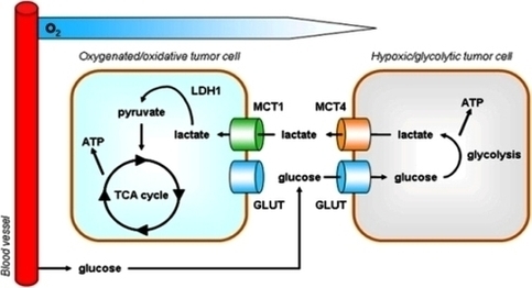
Certainly, the scientific rationale for targeting tumor glycolysis is clearly sound and logical. It is based on the fact that tumor glycolysis is a true signature of cancer cells. A number of drug candidates have been tested mostly pre-clinically with View Diagram Glycolysis Tumor Image
Flu Vaccination Rates Map Image

Aids Cure Would Make Hiv Virus Self Destruct Image Image
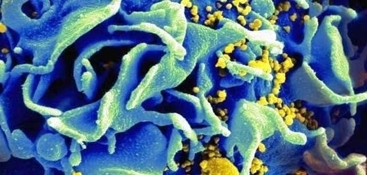
Diagram Illu Dige Tract Image
Looking at pictures of your GI tract can help you to pinpoint where symptoms such as abdominal pain may be coming from. This understanding can also help you to better describe your symptoms to your healthcare provider. Terminal Ileum. The View Diagram Diagram Illu Dige Tract Image
Diagram Easy Plantar Fasciitis Stretches Foot Image
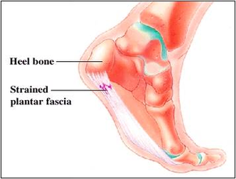
Towel Stretch. To do this stretch, take a towel and put your foot firmly on one end, holding the other in your hands. Pull upwards to stretch all of your toes, stretching out the plantar fascia at the same time. View Diagram Diagram Easy Plantar Fasciitis Stretches Foot Image