
Lower Leg Bones Anatomy
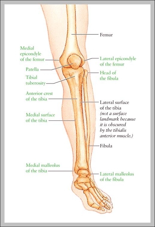
Lower Leg Bones Anatomy: The lower leg contains two main bones: the tibia (shinbone) and fibula. These bones support body weight and connect the knee to the ankle.
Lower Leg Bones Anatomy: The lower leg contains two main bones: the tibia (shinbone) and fibula. These bones support body weight and connect the knee to the ankle.
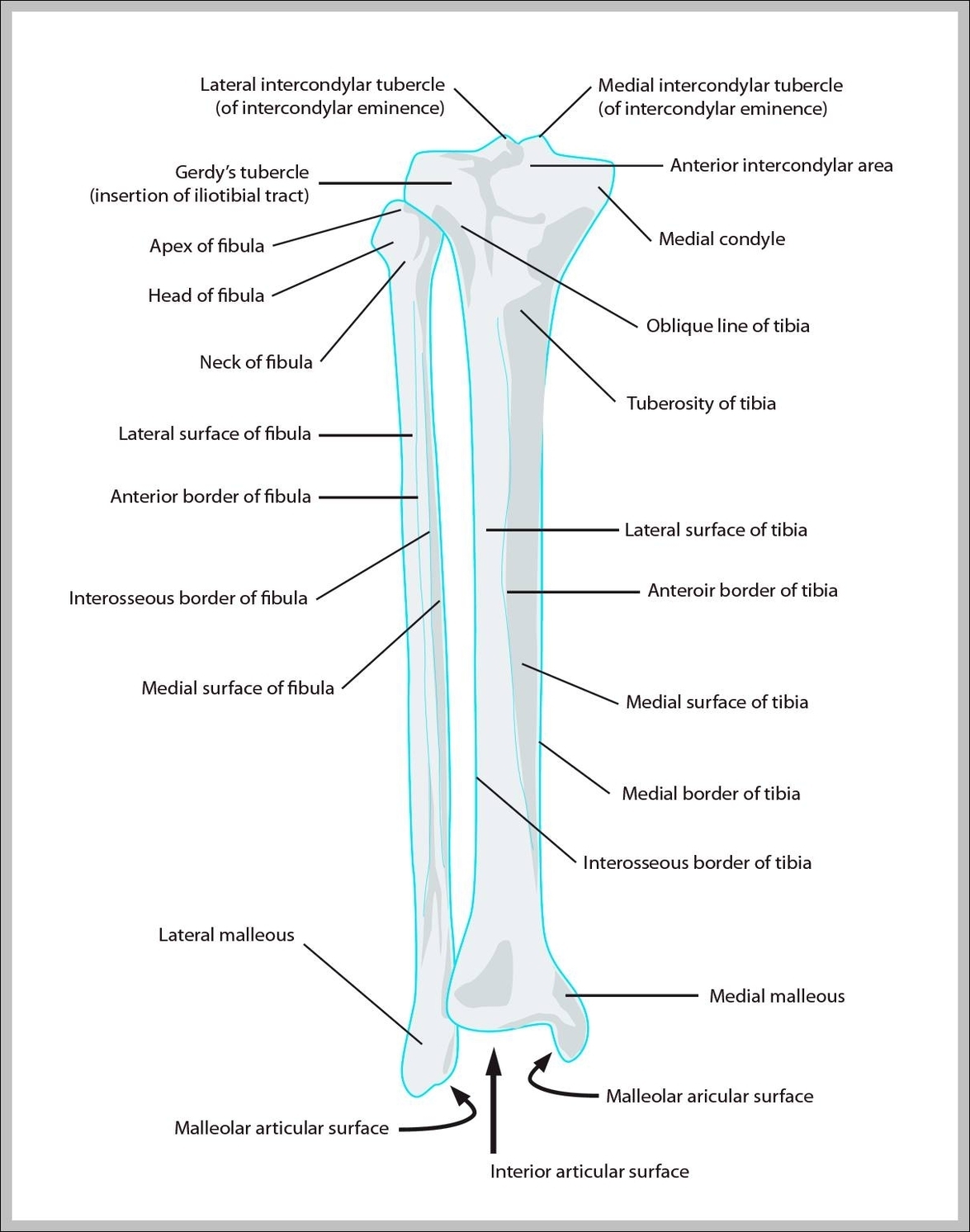
Lower Leg Bones: The lower leg bones include the tibia and fibula, which support body weight and connect the knee to the ankle, essential for walking and bearing loads.
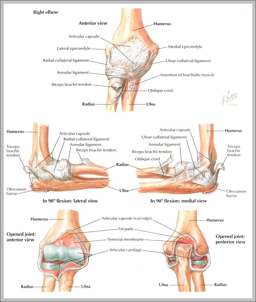
Ligaments Of Elbow: Elbow ligaments, including the ulnar collateral, radial collateral, and annular ligaments, stabilize the joint during arm movement and resist dislocation.
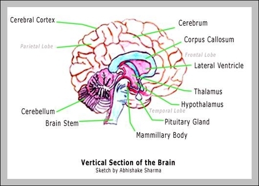
Labeling Parts Of The Brain: A brain labeling diagram identifies structures like the cerebrum, cerebellum, brainstem, and lobes, assisting students in learning brain regions and functions.
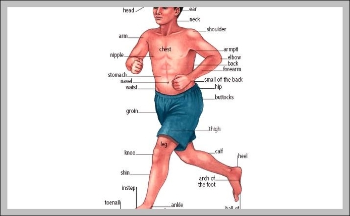
Labeled Body Parts In Spanish: A labeled body parts chart in Spanish provides the names of various body parts in the Spanish language, such as cabeza (head), brazo (arm), and pierna (leg).
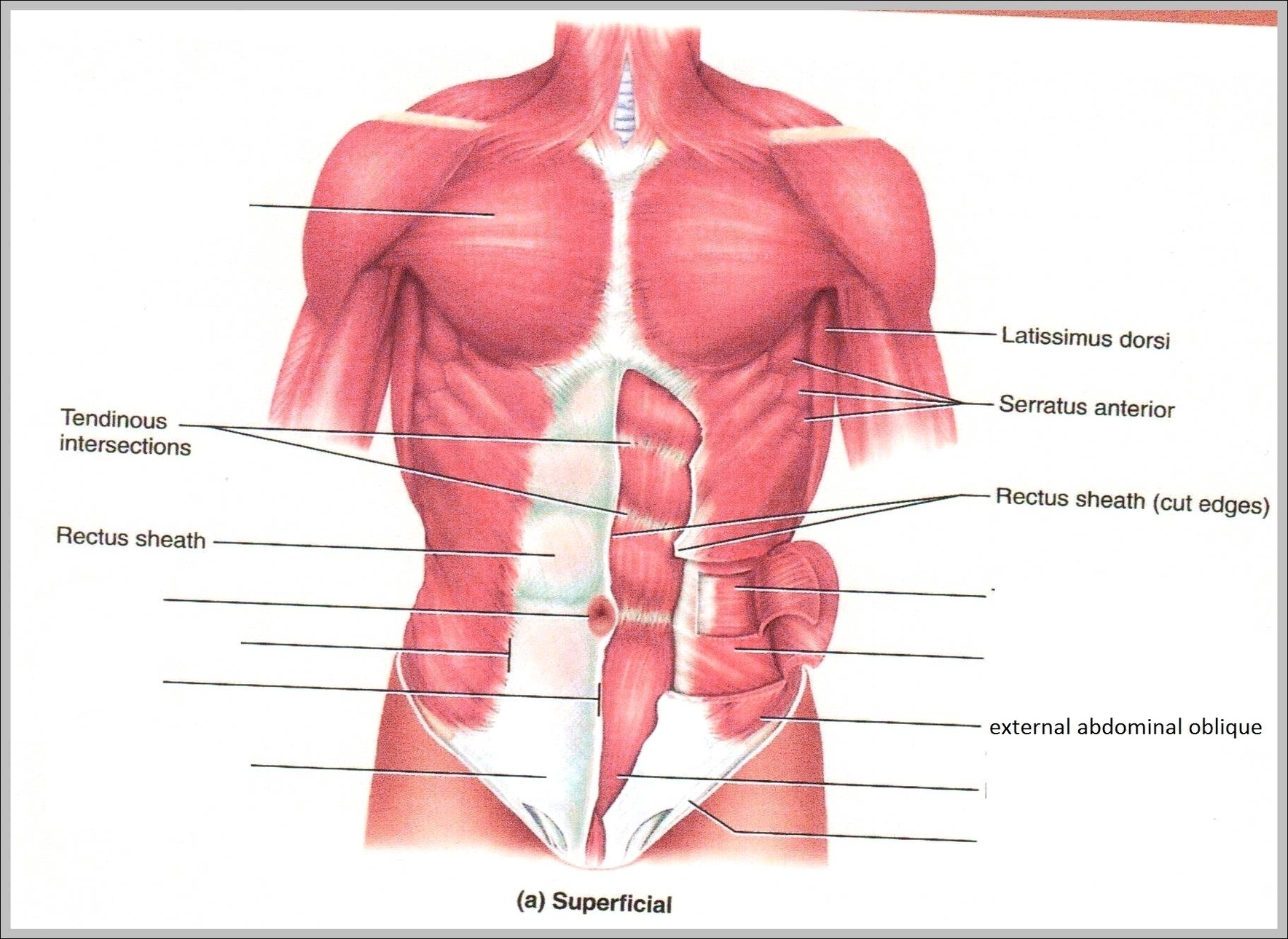
Internal Abdominal Oblique: The internal abdominal oblique muscle lies beneath the external oblique and plays a key role in trunk rotation and lateral flexion. It also aids in compressing the abdominal contents and contributes to core stability.
Inside Your Body Parts: “Inside your body parts” refers to the internal organs and structures of the body that work together to maintain health and perform vital functions, such as the brain, heart, lungs, liver, and kidneys.
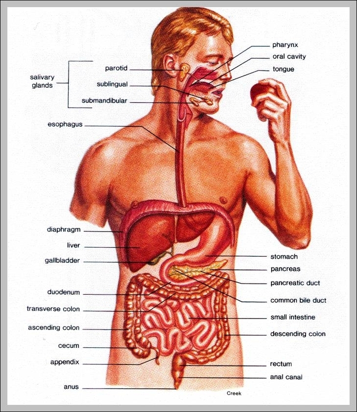
Inside Body Parts: Inside the body are organs such as the brain, lungs, heart, liver, kidneys, intestines, and others, each performing essential tasks to maintain bodily functions.

Inner Parts Of The Body: The inner body includes all internal organs, tissues, and systems such as the heart, lungs, liver, intestines, reproductive organs, and the nervous system.
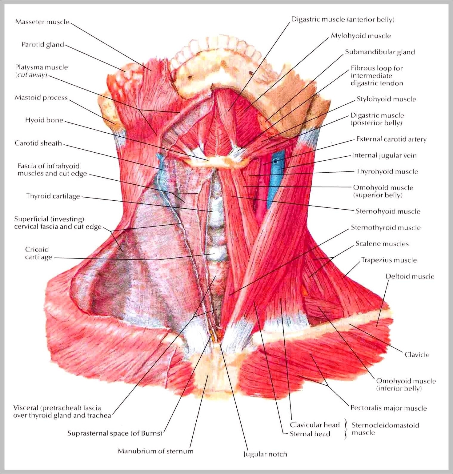
Images Of Neck Muscles: Images of neck muscles show the sternocleidomastoid, trapezius, and scalene muscles, which are important for neck movement and posture.
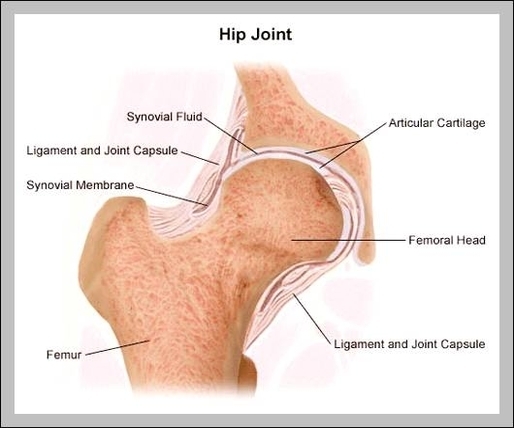
Hip Anatomy Diagram: A hip anatomy diagram highlights the bones and muscles around the hip joint, including the femur, pelvis, and hip flexors, responsible for movement and weight-bearing.
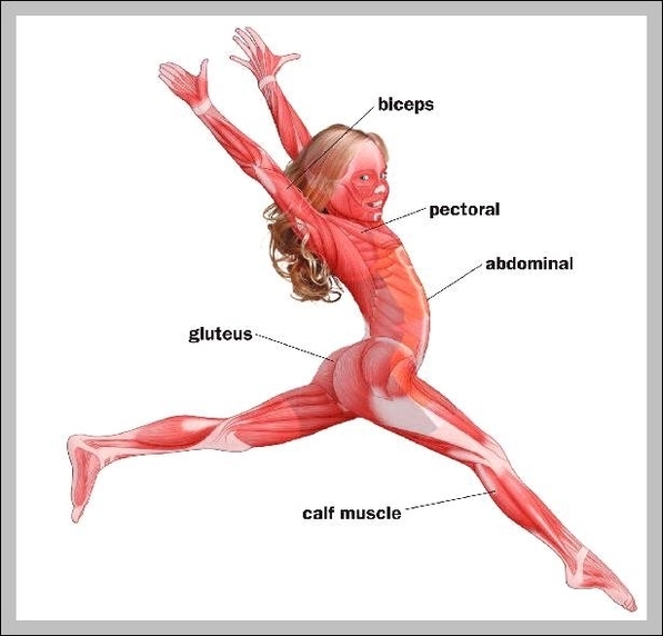
Hip Abductors: Hip abductors, including the gluteus medius and minimus, move the leg away from the bodys midline. They are vital for balance, walking, and preventing hip instability.
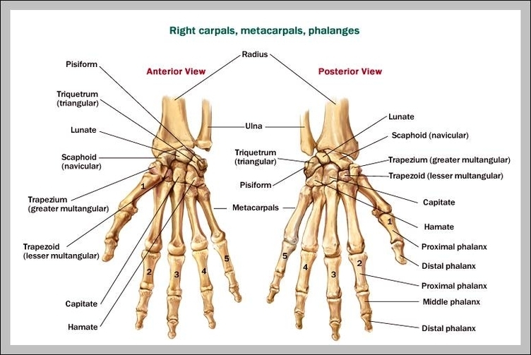
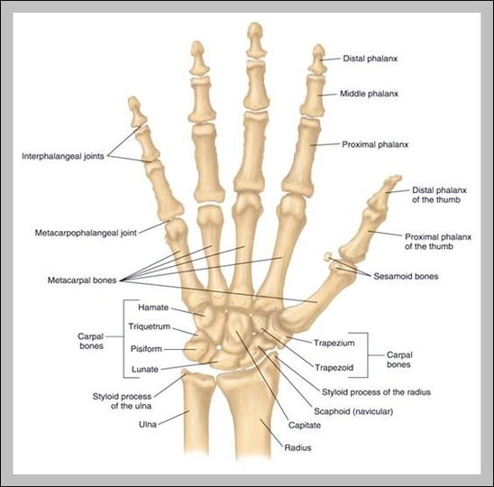
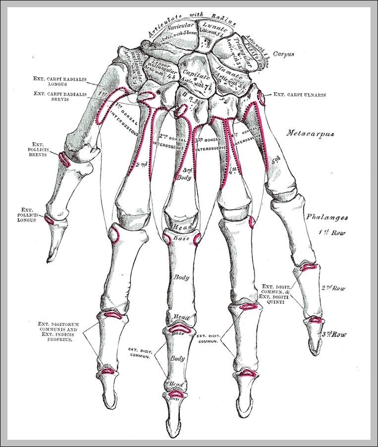
Hand Wrist Bones: The wrist is composed of eight carpal bones that connect the hand to the forearm, while the hand includes metacarpals and phalanges, allowing for a wide range of motion and dexterity.

Hand Ligaments And Tendons: The hand’s ligaments and tendons allow for movement and stability. Ligaments connect bones to bones, while tendons connect muscles to bones, enabling hand functions like grasping and manipulating objects.
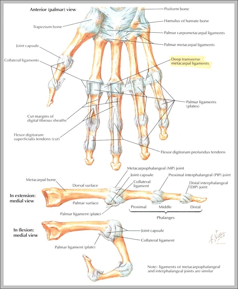
Hand Ligaments: Hand ligaments, such as the collateral ligaments and the flexor retinaculum, provide stability and movement to the wrist, fingers, and thumb, essential for gripping and fine motor skills.
Hand Joints: Hand joints include the wrist (radiocarpal), knuckles (metacarpophalangeal), and finger joints (interphalangeal), providing fine motor control and dexterity.
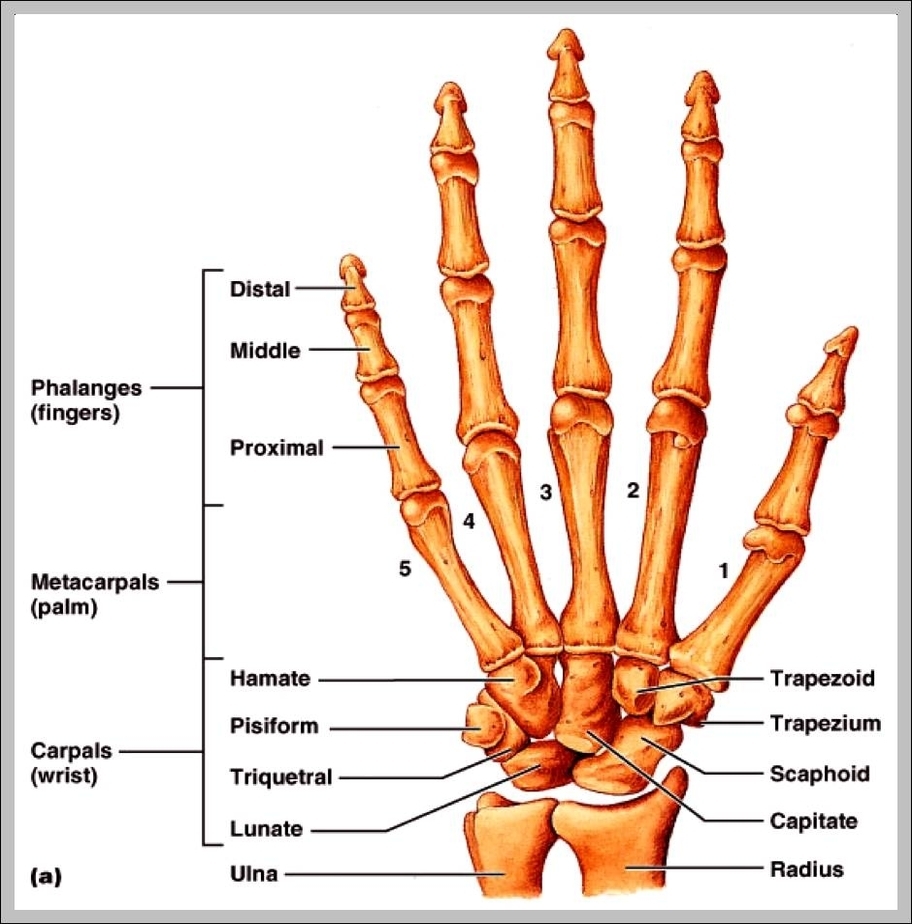
Hand Bones Pictures: Pictures of hand bones show the skeletal structure of the hand, which consists of 27 bones: 8 carpal bones, 5 metacarpals, and 14 phalanges. These bones allow for dexterity and gripping ability.

Hand Bones Anatomy: Hand anatomy includes 27 bonescarpals (wrist), metacarpals (palm), and phalanges (fingers)enabling fine motor skills and grasping.
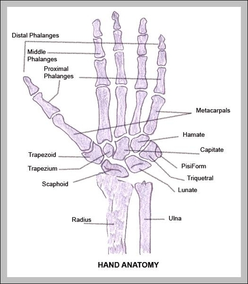
Hand And Wrist Bones Diagram: This diagram displays bones such as the carpals, metacarpals, and phalanges, which together form the complex, flexible structure of the hand and wrist.
Hand Anatomy Picture: A hand anatomy image shows bones, muscles, tendons, and nerves, detailing how intricate structures enable precise movements and grip.