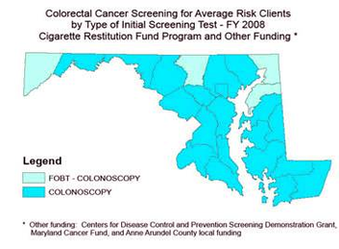
Colon Cancer Screening Types Diagram Image
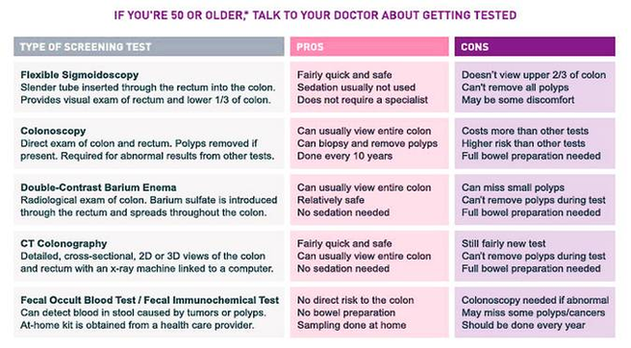
Colorectal Cancer Screening Tests 1 Stool Tests. … 2 Flexible Sigmoidoscopy. … 3 Colonoscopy. … 4 CT Colonography (Virtual Colonoscopy) Computed tomography (CT) colonography, also called a virtual colonoscopy, uses X-rays and computers to produce images of the entire colon, View Diagram Colon Cancer Screening Types Diagram Image